当前位置:
X-MOL 学术
›
Immunol. Cell Biol.
›
论文详情
Our official English website, www.x-mol.net, welcomes your feedback! (Note: you will need to create a separate account there.)
State‐of‐the‐art microscopy to understand islets of Langerhans: what to expect next?
Immunology and Cell Biology ( IF 4 ) Pub Date : 2021-03-05 , DOI: 10.1111/imcb.12450 Pascal de Boer 1 , Ben Ng Giepmans 1
Immunology and Cell Biology ( IF 4 ) Pub Date : 2021-03-05 , DOI: 10.1111/imcb.12450 Pascal de Boer 1 , Ben Ng Giepmans 1
Affiliation
|
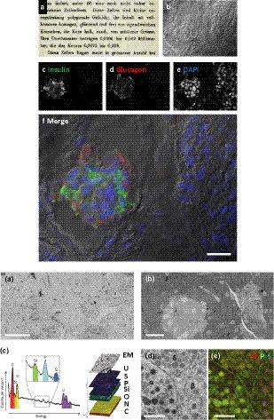
The discovery of Langerhans and microscopic description of islets in the pancreas were crucial steps in the discovery of insulin. Over the past 150 years, many discoveries in islet biology and type 1 diabetes have been made using powerful microscopic techniques. In the past decade, combination of new probes, animal and tissue models, application of new biosensors and automation of light and electron microscopic methods and other (sub)cellular imaging modalities have proven their potential in understanding the beta cell under (patho)physiological conditions. The imaging evolution, from fluorescent jellyfish to real‐time intravital functional imaging, the revolution in automation and data handling and the increased resolving power of analytical imaging techniques are now converging. Here, we review innovative approaches that address islet biology from new angles by studying cells and molecules at high spatiotemporal resolution and in live models. Broad implementation of these cellular imaging techniques will shed new light on cause/consequence of (mal)function in islets of Langerhans in the years to come.
中文翻译:

了解朗格汉斯胰岛的最先进显微镜:接下来会发生什么?
朗格汉斯的发现和胰腺中胰岛的微观描述是发现胰岛素的关键步骤。在过去的 150 年里,胰岛生物学和 1 型糖尿病方面的许多发现都是使用强大的显微技术取得的。在过去的十年中,新探针、动物和组织模型的结合、新生物传感器的应用以及光和电子显微方法的自动化以及其他(亚)细胞成像模式已经证明了它们在(病理)生理条件下理解 β 细胞方面的潜力. 成像进化,从荧光水母到实时活体功能成像,自动化和数据处理的革命以及分析成像技术分辨率的提高正在融合。这里,我们通过在高时空分辨率和活体模型中研究细胞和分子,从新角度回顾了解决胰岛生物学的创新方法。未来几年,这些细胞成像技术的广泛应用将为朗格汉斯岛功能(故障)的原因/后果提供新的线索。
更新日期:2021-05-11
中文翻译:
了解朗格汉斯胰岛的最先进显微镜:接下来会发生什么?
朗格汉斯的发现和胰腺中胰岛的微观描述是发现胰岛素的关键步骤。在过去的 150 年里,胰岛生物学和 1 型糖尿病方面的许多发现都是使用强大的显微技术取得的。在过去的十年中,新探针、动物和组织模型的结合、新生物传感器的应用以及光和电子显微方法的自动化以及其他(亚)细胞成像模式已经证明了它们在(病理)生理条件下理解 β 细胞方面的潜力. 成像进化,从荧光水母到实时活体功能成像,自动化和数据处理的革命以及分析成像技术分辨率的提高正在融合。这里,我们通过在高时空分辨率和活体模型中研究细胞和分子,从新角度回顾了解决胰岛生物学的创新方法。未来几年,这些细胞成像技术的广泛应用将为朗格汉斯岛功能(故障)的原因/后果提供新的线索。


 京公网安备 11010802027423号