Applied Microbiology and Biotechnology ( IF 5 ) Pub Date : 2021-02-10 , DOI: 10.1007/s00253-021-11098-0 Zui Fujimoto , Naomi Kishine , Koji Teramoto , Sosyu Tsutsui , Satoshi Kaneko
|
Abstract
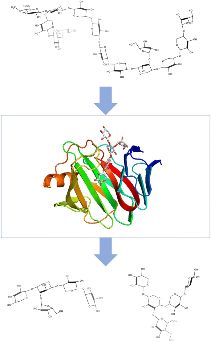
Although many xylanases have been studied, many of the characteristics of xylanases toward branches in xylan remain unclear. In this study, the substrate specificity of a GH11 xylanase from Streptomyces olivaceoviridis E-86 (SoXyn11B) was elucidated based on its three-dimensional structure. Subsite mapping suggests that SoXyn11B has seven subsites (four subsites on the – side and three subsites on the + side), and it is one longer than the GH10 xylanase from S. olivaceoviridis (SoXyn10A). SoXyn11B has no affinity for the subsites at either end of the scissile glycosidic bond, and the sugar-binding energy at subsite – 2 was the highest, followed by subsite + 2. These properties were very similar to those of SoXyn10A. In contrast, SoXyn11B produced different branched oligosaccharides from bagasse compared with those of SoXyn10A. These branched oligosaccharides were identified as O-β-D-xylopyranosyl-(1→4)-[O-α-L-arabinofuranosyl-(1→3)]-O-β-D-xylopyranosyl-(1→4)-β-D-xylopyranosyl-(1→4)-β-D-xylopyranose (Ara3Xyl4) and O-β-D-xylopyranosyl-(1→4)-[O-4-O-methyl-α-D-glucuronopyranosyl-(l→2)]-β-D-xylopyranosyl-(1→4)-β-D-xylopyranosyl-(1→4)-β-D-xylopyranose (MeGlcA3Xyl4) by nuclear magnetic resonance (NMR) and electrospray ionization mass spectrometry (ESI-MS) and confirmed by crystal structure analysis of SoXyn11B in complex with these branched xylooligosaccharides. SoXyn11B has a β-jerryroll fold structure, and the catalytic cleft is located on the inner β-sheet of the fold. The ligand-binding structures revealed seven subsites of SoXyn11B. The 2- and 3-hydroxy groups of xylose at the subsites + 3, + 2, and – 3 face outwards, and an arabinose or a glucuronic acid side chain can be linked to these positions. These subsite structures appear to cause the limited substrate specificity of SoXyn11B for branched xylooligosaccharides.
Key points
• Crystal structure of family 11 β-xylanase from Streptomyces olivaceoviridis was determined.
• Topology of substrate-binding cleft of family 11 β-xylanase from Streptomyces olivaceoviridis was characterized.
• Mode of action of family 11 β-xylanase from Streptomyces olivaceoviridis for substitutions in xylan was elucidated.
Graphical abstract
中文翻译:
寡链链霉菌E-86中GH11木聚糖酶的基于结构的底物特异性分析
摘要
尽管已经研究了许多木聚糖酶,但是木聚糖酶对木聚糖分支的许多特征仍然不清楚。在这项研究中,基于寡聚链霉菌E-86(SoXyn11B)的三维结构,阐明了其GH11木聚糖酶的底物特异性。子站点映射表明,SoXyn11B具有七个子站点(–侧为四个子站点,+端为三个子站点),它比寡糖链球菌的GH10木聚糖酶长一个。(SoXyn10A)。SoXyn11B对易裂糖苷键两端的亚位无亲和力,并且亚位处的糖结合能最高,为2,其次为亚位+2。这些特性与SoXyn10A非常相似。相反,与SoXyn10A相比,SoXyn11B从甘蔗渣中产生了不同的分支寡糖。这些支链低聚糖被鉴定为O -β-D-吡喃喃糖基-(1→4)-[ O -α-L-阿拉伯呋喃糖基-(1→3)]- O -β-D-吡喃吡喃糖基-(1→4)- β-D-吡喃喃糖基-(1→4)-β-D-吡喃吡喃糖(Ara 3 Xyl 4)和O - β-D-吡喃吡喃糖基-(1→4)-[ O -4- O-甲基-α-D-葡糖醛酸吡喃糖基-(1→2)]-β-D-吡喃吡喃糖基-(1→4)-β-D-吡喃吡喃糖基-(1→4)-β-D-吡喃吡喃糖(MeGlcA 3 Xyl 4通过核磁共振(NMR)和电喷雾电离质谱(ESI-MS),并通过SoXyn11B与这些支链低聚木糖复合的晶体结构分析得到证实。SoXyn11B具有β-jerryroll折叠结构,催化裂隙位于折叠的内部β-折叠上。配体结合结构揭示了SoXyn11B的七个亚位点。木糖的+ 3,+ 2和–3亚基上的木糖的2-和3-羟基朝外,阿拉伯糖或葡糖醛酸侧链可以连接到这些位置。这些亚位点结构似乎导致SoXyn11B对支链低聚木糖的底物特异性有限。
关键点
•确定了来自Streptomyces olivaceoviridis的11族β-木聚糖酶的晶体结构。
•表征了来自Streptomyces olivaceoviridis的11族β-木聚糖酶的底物结合裂隙的拓扑结构。
•阐明了来自Streptomyces olivaceoviridis的11族β-木聚糖酶替代木聚糖的作用方式。


 京公网安备 11010802027423号