Spectrochimica Acta Part A: Molecular and Biomolecular Spectroscopy ( IF 4.4 ) Pub Date : 2021-02-05 , DOI: 10.1016/j.saa.2021.119552 Hamed.M. Al-Saidi , Salman.S. Alharthi
|
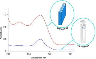
In this paper, dispersive liquid–liquid microextraction (DLLME), long optical path microcells, and a selective chromogenic reagent were employed to improve the analytical efficiency of cobalt determination by spectrophotometry. The methodology proposed in the present study is based upon the microextraction of a cobalt(II) complex with 1-[4-[(2-hydroxynaphthalen-1-yl)methylideneamino] phenyl]ethanone (HNE) by DLLME and measurement of the absorbance of the sedimented phase using a microcell with an optical path length of 50 mm (Microcell–50). DLLME was performed using a binary mixture containing 900 μL of methanol as a dispersing solvent and 400 μL of CHCl3 (extraction solvent) at pH 6–8 adjusted by a mixture of HCl and NaOH. The electronic spectrum of the dark brown complex recorded in the sedimented phase using Microcell–50 shows a well-defined peak at λmax 324 ± 3 nm with a molar absorptivity of 1.08 × 10⁶ M−1 cm−1. Cobalt was monitored at a detection limit (LOD) of 0.08 μg L–1 and in the linear concentration range of 0.45–10 μg L–1, while the limit of quantitation (LOQ), relative standard deviation (RSD), and the enhancement factor (EF) were 0.264, 1.6 μgL–1, and 223, respectively. Our method was evaluated by determining cobalt in certified reference materials and experimental samples, and the results were compared with ICP–MS measurements. Moreover, the chemical structure of the [Co(C38H28O2N)2] complex was suggested through using different characterization techniques such as Fourier transform infrared spectroscopy (FT-IR), scanning electron microscopy (SEM), energy-dispersive X-ray spectroscopy (EDX), thermal analysis, and powder X-ray diffraction.
中文翻译:
使用分散液液微萃取和具有长光程的微池提高水和药物制剂中钴的分光光度估算效率
在本文中,采用分散液液微萃取(DLLME),长光程微池和选择性生色试剂来提高分光光度法测定钴的分析效率。本研究中提出的方法是基于通过DLLME微萃取钴(II)与1- [4-[(2-羟基萘-1-基)亚甲基氨基]苯基]乙酮(HNE)的配合物并测量吸光度使用光程长度为50 mm的微池(Microcell–50)对沉淀相进行分析。使用包含900μL甲醇作为分散溶剂和400μLCHCl 3的二元混合物进行DLLME(萃取溶剂),pH为6-8,由HCl和NaOH的混合物调节。使用Microcell–50在沉积相中记录的暗褐色络合物的电子光谱在λmax 324±3 nm处有一个明确定义的峰,摩尔吸收率为1.08×10 6 M -1 cm -1。钴在检测限为0.08微克L(LOD)进行了监测-1和在0.45-10微克L中的线性浓度范围-1,而定量限(LOQ)的限制,相对标准偏差(RSD),和增强因子(EF)为0.264,1.6μgL –1,和223。通过测定经认证的参考物质和实验样品中的钴对我们的方法进行了评估,并将结果与ICP-MS测量结果进行了比较。此外,[Co(C 38 H 28 O 2 N)2 ]配合物的化学结构是通过使用不同的表征技术,例如傅立叶变换红外光谱(FT-IR),扫描电子显微镜(SEM),能量色散,提出的X射线光谱(EDX),热分析和粉末X射线衍射。


 京公网安备 11010802027423号