当前位置:
X-MOL 学术
›
Bioconjugate Chem.
›
论文详情
Our official English website, www.x-mol.net, welcomes your feedback! (Note: you will need to create a separate account there.)
ImmunoPET of CD146 in Orthotopic and Metastatic Breast Cancer Models
Bioconjugate Chemistry ( IF 4.7 ) Pub Date : 2021-01-21 , DOI: 10.1021/acs.bioconjchem.0c00649 Cuicui Li 1 , Lei Kang 1, 2 , Kevin Fan 2 , Carolina A Ferreira 3 , Kaelyn V Becker 2 , Nan Huo 4 , Hanxiao Liu 5 , Yunan Yang 2 , Jonathan W Engle 2 , Rongfu Wang 1 , Xiaojie Xu 4 , Dawei Jiang 2, 6 , Weibo Cai 2, 3
Bioconjugate Chemistry ( IF 4.7 ) Pub Date : 2021-01-21 , DOI: 10.1021/acs.bioconjchem.0c00649 Cuicui Li 1 , Lei Kang 1, 2 , Kevin Fan 2 , Carolina A Ferreira 3 , Kaelyn V Becker 2 , Nan Huo 4 , Hanxiao Liu 5 , Yunan Yang 2 , Jonathan W Engle 2 , Rongfu Wang 1 , Xiaojie Xu 4 , Dawei Jiang 2, 6 , Weibo Cai 2, 3
Affiliation

|
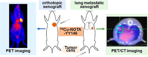
The overexpression of CD146 in breast cancer is considered a hallmark of tumor progression and metastasis, particularly in triple negative breast cancer. Aimed at imaging differential CD146 expressions in breast cancer, a noninvasive method for predictive prognosis and diagnosis was investigated using a 64Cu-labeled CD146-specific monoclonal antibody, YY146. CD146 expression was screened in human breast cancer cell lines using Western blotting. Binding ability was evaluated using flow cytometry and immunofluorescent staining. YY146 was conjugated with 1,4,7-triazacyclononane-triacetic acid (NOTA) and radiolabeled with 64Cu following standard procedures. Serial PET or PET/CT imaging was performed in orthotopic and metastatic breast cancer tumor models. Biodistribution was performed after the final time point of imaging. Finally, tissue immunofluorescent staining and hematoxylin and eosin (H&E) staining were performed on tumor tissues. The MDA-MB-435 cell line showed the highest CD146 expression level, whereas MCF-7 had the lowest level at the cellular level. ImmunoPET showed that MDA-MB-435 orthotopic tumors had high and clear radioactive accumulation after the administration of 64Cu-NOTA-YY146. The tumor uptake of 64Cu-NOTA-YY146 in MDA-MB-435 was significantly higher than that in MCF-7 and nonspecific IgG control groups (P < 0.01). Biodistribution verified the PET imaging results. For metastatic models, 64Cu-NOTA-YY146 allowed for the visualization of high radioactivity accumulation in metastatic MDA-MB-435 tumors, which was confirmed by ex vivo biodistribution of lung tissues. H&E staining proved the successful building of metastatic tumor models. Immunofluorescent staining verified the differential expression of CD146 in orthotopic tumors. Therefore, 64Cu-NOTA-YY146 could be used as an immunoPET probe to visualize CD146 in the breast cancer model and is potentially useful for cancer diagnosis, prognosis prediction, and monitoring therapeutic response.
中文翻译:

CD146 在原位和转移性乳腺癌模型中的免疫 PET
CD146 在乳腺癌中的过表达被认为是肿瘤进展和转移的标志,特别是在三阴性乳腺癌中。针对乳腺癌中 CD146 差异表达的成像,使用64 Cu 标记的 CD146 特异性单克隆抗体 YY146 研究了一种预测预后和诊断的无创方法。使用蛋白质印迹法在人乳腺癌细胞系中筛选 CD146 表达。使用流式细胞术和免疫荧光染色评估结合能力。YY146 与 1,4,7-三氮杂环壬烷-三乙酸 (NOTA) 结合并用64进行放射性标记铜遵循标准程序。在原位和转移性乳腺癌肿瘤模型中进行系列 PET 或 PET/CT 成像。在成像的最后时间点之后进行生物分布。最后,对肿瘤组织进行组织免疫荧光染色和苏木精和伊红(H&E)染色。MDA-MB-435 细胞系的 CD146 表达水平最高,而 MCF-7 在细胞水平上的表达水平最低。ImmunoPET显示MDA-MB-435原位肿瘤在给予64 Cu-NOTA-YY146后具有高且明显的放射性蓄积。MDA-MB-435对64 Cu-NOTA-YY146的肿瘤摄取显着高于MCF-7和非特异性IgG对照组(P< 0.01)。生物分布验证了 PET 成像结果。对于转移模型,64 Cu-NOTA-YY146 允许在转移性 MDA-MB-435 肿瘤中显示高放射性积累,这已通过肺组织的离体生物分布得到证实。H&E染色证明转移性肿瘤模型的成功构建。免疫荧光染色验证了CD146在原位肿瘤中的差异表达。因此,64 Cu-NOTA-YY146 可用作免疫PET 探针以可视化乳腺癌模型中的CD146,并可能用于癌症诊断、预后预测和监测治疗反应。
更新日期:2021-01-21
中文翻译:

CD146 在原位和转移性乳腺癌模型中的免疫 PET
CD146 在乳腺癌中的过表达被认为是肿瘤进展和转移的标志,特别是在三阴性乳腺癌中。针对乳腺癌中 CD146 差异表达的成像,使用64 Cu 标记的 CD146 特异性单克隆抗体 YY146 研究了一种预测预后和诊断的无创方法。使用蛋白质印迹法在人乳腺癌细胞系中筛选 CD146 表达。使用流式细胞术和免疫荧光染色评估结合能力。YY146 与 1,4,7-三氮杂环壬烷-三乙酸 (NOTA) 结合并用64进行放射性标记铜遵循标准程序。在原位和转移性乳腺癌肿瘤模型中进行系列 PET 或 PET/CT 成像。在成像的最后时间点之后进行生物分布。最后,对肿瘤组织进行组织免疫荧光染色和苏木精和伊红(H&E)染色。MDA-MB-435 细胞系的 CD146 表达水平最高,而 MCF-7 在细胞水平上的表达水平最低。ImmunoPET显示MDA-MB-435原位肿瘤在给予64 Cu-NOTA-YY146后具有高且明显的放射性蓄积。MDA-MB-435对64 Cu-NOTA-YY146的肿瘤摄取显着高于MCF-7和非特异性IgG对照组(P< 0.01)。生物分布验证了 PET 成像结果。对于转移模型,64 Cu-NOTA-YY146 允许在转移性 MDA-MB-435 肿瘤中显示高放射性积累,这已通过肺组织的离体生物分布得到证实。H&E染色证明转移性肿瘤模型的成功构建。免疫荧光染色验证了CD146在原位肿瘤中的差异表达。因此,64 Cu-NOTA-YY146 可用作免疫PET 探针以可视化乳腺癌模型中的CD146,并可能用于癌症诊断、预后预测和监测治疗反应。


 京公网安备 11010802027423号