Our official English website, www.x-mol.net, welcomes your feedback! (Note: you will need to create a separate account there.)
The influence of the morphology of titania and hydroxyapatite on the proliferation and osteogenic differentiation of human mesenchymal stem cells
RSC Advances ( IF 3.9 ) Pub Date : 2021-1-20 , DOI: 10.1039/d0ra08271f Yauheni U Kuvyrkou 1, 2 , Nadzeya Brezhneva 3 , Ekaterina V Skorb 4 , Sviatlana A Ulasevich 4
RSC Advances ( IF 3.9 ) Pub Date : 2021-1-20 , DOI: 10.1039/d0ra08271f Yauheni U Kuvyrkou 1, 2 , Nadzeya Brezhneva 3 , Ekaterina V Skorb 4 , Sviatlana A Ulasevich 4
Affiliation
|
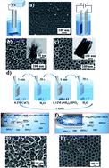
Herein, the proliferation and osteogenic potential of human mesenchymal stem cells (hMSCs) on the disordered and ordered porous morphology of the titania surface and titania surface modified by hydroxyapatite (HA) are compared for the first time. In 5 days, the MTT-assay showed that the ordered porous morphology of electrochemically fabricated titania nanotubes (TNT) and TNT with chemically deposited hydroxyapatite (TNT–HA) was favorable for stem cell proliferation. In 14 days, RT-qPCR demonstrated that the disordered porous morphology of the sonochemically produced titania mesoporous surface (TMS) and TMS modified by the chemical deposition of HA (TMS–HA) led to the differentiation of hMSCs into the osteogenic direction in the absence of osteogenic inductors. These results originate from the mechanism of mechanotransduction, which sheds a light on the interaction of mesenchymal stem cells with the porous interface through focal adhesion, regulating the expression of genes determining stem cell self-renewal and osteogenic differentiation. The strong focal adhesion of hMSCs adjusted by the disordered TMS and TMS–HA is enough to induce osteogenic differentiation with the delay of cellular self-renewal. The weak focal adhesion of hMSCs tuned by the ordered TNT and TNT–HA affects only cellular self-renewal. The present research makes a new contribution to nanomedicine and engineering of porous implant interfaces for the replacement of bone injuries.
中文翻译:

二氧化钛和羟基磷灰石形态对人间充质干细胞增殖和成骨分化的影响
在此,首次比较了人间充质干细胞 (hMSCs) 在二氧化钛表面和羟基磷灰石 (HA) 修饰的二氧化钛表面无序有序多孔形态上的增殖和成骨潜力。在 5 天内,MTT 分析表明,电化学制造的二氧化钛纳米管 (TNT) 和具有化学沉积羟基磷灰石 (TNT-HA) 的 TNT 的有序多孔形态有利于干细胞增殖。在 14 天内,RT-qPCR 证明声化学产生的二氧化钛介孔表面 (TMS) 和经 HA 化学沉积修饰的 TMS (TMS-HA) 的无序多孔形态导致 hMSC 在不存在的情况下向成骨方向分化成骨诱导剂。这些结果源于机械转导机制,这揭示了间充质干细胞通过粘着斑与多孔界面的相互作用,调节决定干细胞自我更新和成骨分化的基因的表达。通过无序的 TMS 和 TMS-HA 调节的 hMSCs 的强粘着斑足以诱导成骨分化,延迟细胞自我更新。由有序 TNT 和 TNT-HA 调节的 hMSCs 的弱粘着斑仅影响细胞自我更新。本研究为纳米医学和多孔植入物界面工程以替代骨损伤做出了新的贡献。通过无序的 TMS 和 TMS-HA 调节的 hMSCs 的强粘着斑足以诱导成骨分化,延迟细胞自我更新。由有序 TNT 和 TNT-HA 调节的 hMSCs 的弱粘着斑仅影响细胞自我更新。本研究为纳米医学和多孔植入物界面工程以替代骨损伤做出了新的贡献。通过无序的 TMS 和 TMS-HA 调节的 hMSCs 的强粘着斑足以诱导成骨分化,延迟细胞自我更新。由有序 TNT 和 TNT-HA 调节的 hMSCs 的弱粘着斑仅影响细胞自我更新。本研究为纳米医学和多孔植入物界面工程以替代骨损伤做出了新的贡献。
更新日期:2021-01-20
中文翻译:
二氧化钛和羟基磷灰石形态对人间充质干细胞增殖和成骨分化的影响
在此,首次比较了人间充质干细胞 (hMSCs) 在二氧化钛表面和羟基磷灰石 (HA) 修饰的二氧化钛表面无序有序多孔形态上的增殖和成骨潜力。在 5 天内,MTT 分析表明,电化学制造的二氧化钛纳米管 (TNT) 和具有化学沉积羟基磷灰石 (TNT-HA) 的 TNT 的有序多孔形态有利于干细胞增殖。在 14 天内,RT-qPCR 证明声化学产生的二氧化钛介孔表面 (TMS) 和经 HA 化学沉积修饰的 TMS (TMS-HA) 的无序多孔形态导致 hMSC 在不存在的情况下向成骨方向分化成骨诱导剂。这些结果源于机械转导机制,这揭示了间充质干细胞通过粘着斑与多孔界面的相互作用,调节决定干细胞自我更新和成骨分化的基因的表达。通过无序的 TMS 和 TMS-HA 调节的 hMSCs 的强粘着斑足以诱导成骨分化,延迟细胞自我更新。由有序 TNT 和 TNT-HA 调节的 hMSCs 的弱粘着斑仅影响细胞自我更新。本研究为纳米医学和多孔植入物界面工程以替代骨损伤做出了新的贡献。通过无序的 TMS 和 TMS-HA 调节的 hMSCs 的强粘着斑足以诱导成骨分化,延迟细胞自我更新。由有序 TNT 和 TNT-HA 调节的 hMSCs 的弱粘着斑仅影响细胞自我更新。本研究为纳米医学和多孔植入物界面工程以替代骨损伤做出了新的贡献。通过无序的 TMS 和 TMS-HA 调节的 hMSCs 的强粘着斑足以诱导成骨分化,延迟细胞自我更新。由有序 TNT 和 TNT-HA 调节的 hMSCs 的弱粘着斑仅影响细胞自我更新。本研究为纳米医学和多孔植入物界面工程以替代骨损伤做出了新的贡献。


 京公网安备 11010802027423号