Journal of Neuroradiology ( IF 3.5 ) Pub Date : 2021-01-19 , DOI: 10.1016/j.neurad.2021.01.002 Liuji Guo 1 , Xiaodan Li 1 , Haimei Cao 1 , Jun Hua 2 , Yingjie Mei 3 , Jay J Pillai 4 , Yuankui Wu 1
|
Purpose
The aim of the study is to assess the diagnostic performance of inflow-based vascular-space-occupancy (iVASO) MR imaging for differentiating glioblastomas (grade IV, GBM) and lower-grade diffuse gliomas (grade II and III, LGG) and its potential to predict IDH mutation status.
Methods
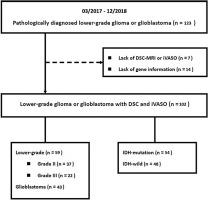
One hundred and two patients with diffuse cerebral glioma (56 males; median age, 43.5 years) underwent iVASO and dynamic susceptibility contrast (DSC) MR imaging. The iVASO-derived arteriolar cerebral blood volume (CBVa), relative CBVa (rCBVa), and the DSC-derived relative cerebral blood volume (rCBV) were obtained, and these measurements were compared between the GBM group (n = 43) and the LGG group (n = 59) and between the IDH-mutation group (n = 54) and the IDH-wild group (n = 48).
Results
Significant correlation was observed between rCBV and CBVa (P < 0.001) or rCBVa (P < 0.001). Both CBVa (P < 0.001) and rCBVa (P < 0.001) were higher in the GBM group. Both CBVa (P < 0.001) and rCBVa (P < 0.001) were lower in the IDH-mutation group compared to the IDH-wild group. Receiver operating characteristic analyses showed the area under curve (AUC) of 0.95 with CBVa and 0.97 with rCBVa in differentiating GBM from LGG. The AUCs were 0.82 and 0.85 for CBVa and rCBVa in predicting IDH gene status, respectively, which were lower than that of rCBV (AUC = 0.90). Combined rCBV and rCBVa significantly improved the diagnostic performance (AUC = 0.95).
Conclusions
iVASO MR imaging has the potential to predict IDH mutation and grade in glioma.
中文翻译:
基于流入的血管占位(iVASO)可能预测弥漫性脑胶质瘤的 IDH 突变状态和肿瘤分级
目的
本研究的目的是评估基于流入的血管占位 (iVASO) MR 成像对区分胶质母细胞瘤(IV 级,GBM)和低级别弥漫性胶质瘤(II 级和 III 级,LGG)的诊断性能及其预测 IDH 突变状态的潜力。
方法
102 名弥漫性脑胶质瘤患者(56 名男性;中位年龄 43.5 岁)接受了 iVASO 和动态磁敏对比 (DSC) MR 成像。获得了 iVASO 衍生的小动脉脑血容量 (CBVa)、相对 CBVa (rCBVa) 和 DSC 衍生的相对脑血容量 (rCBV),并将这些测量值在 GBM 组 (n = 43) 和 LGG 之间进行了比较组 (n = 59) 和 IDH 突变组 (n = 54) 和 IDH-野生组 (n = 48) 之间。
结果
rCBV 与 CBVa (P < 0.001) 或 rCBVa (P < 0.001) 之间存在显着相关性。GBM 组的 CBVa (P < 0.001) 和 rCBVa (P < 0.001) 均较高。与 IDH 野生组相比,IDH 突变组的 CBVa (P < 0.001) 和 rCBVa (P < 0.001) 均较低。接受者操作特征分析显示,在区分 GBM 和 LGG 时,CBVa 和 rCBVa 的曲线下面积 (AUC) 分别为 0.95 和 0.97。CBVa 和 rCBVa 预测 IDH 基因状态的 AUC 分别为 0.82 和 0.85,低于 rCBV(AUC = 0.90)。结合 rCBV 和 rCBVa 显着提高了诊断性能(AUC = 0.95)。
结论
iVASO MR 成像具有预测胶质瘤 IDH 突变和分级的潜力。


 京公网安备 11010802027423号