Infrared Physics & Technology ( IF 3.3 ) Pub Date : 2021-01-18 , DOI: 10.1016/j.infrared.2020.103611 Junsoo Lee , Jihun Ryu , Sangyeob Han , Naresh Kumar Ravichandran , Daewoon Seong , Jaeyul Lee , Ruchire Eranga Wijesinghe , Pilun Kim , Seung-Yeol Lee , Hee-Young Jung , Mansik Jeon , Kwang Shik Choi , Jeehyun Kim
|
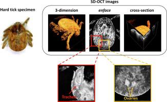
Ixodidae tick, also known as a hard tick, is one of the major vectors of various tick-borne diseases. Studying its anatomy is the fundamental approach for diverse acarological studies and the key to understanding tick morphology. However, the conventional methods of observing internal organs rely primarily on dissection, which damages specimens irrecoverably. In this study, we developed an optical coherence tomography (OCT) system to non-invasively investigate the morphological characteristics of the hard tick. Herein, OCT imaging was conducted by the developed spectral-domain OCT (SD-OCT) system with two different objective lenses. The developed system provides an axial resolution (in the air) of 6.2 µm and a maximum lateral resolution of 2.46 µm as an objective lens with a high numerical aperture (NA) and 10× magnification was employed. Using the developed SD-OCT system, internal organs of tick specimens, such as salivary glands, midgut, genital orifice, and ovary, were identified without inflicting damage. The study suggests the feasibility of the optical coherence imaging for the acarological study of the fundamental morphological inspection and for possible future studies, such as verifying the potential morphological differences among virus transmitted hard tick specimens.
中文翻译:
光谱域光学相干断层扫描技术识别硬tick体内的器官
x科tick科,也称为硬tick科,是各种tick传播疾病的主要媒介之一。研究其解剖结构是进行各种航空学研究的基本方法,也是了解壁虱形态的关键。然而,传统的观察内部器官的方法主要依靠解剖,这会无法修复地破坏标本。在这项研究中,我们开发了光学相干断层扫描(OCT)系统,以非侵入性的方式研究硬壁虱的形态特征。在本文中,OCT成像是通过开发的具有两个不同物镜的光谱域OCT(SD-OCT)系统进行的。所开发的系统可提供6.2 µm的轴向分辨率(空气中)和2.46 µm的最大横向分辨率,这是因为采用了具有高数值孔径(NA)和10倍放大率的物镜。使用已开发的SD-OCT系统,可以在不造成损害的情况下识别壁虱标本的内部器官,例如唾液腺,中肠,生殖孔和卵巢。该研究表明光学相干成像技术在进行基本形态学检查的航空学研究以及可能的未来研究中的可行性,例如验证病毒传播的硬tick标本之间潜在的形态学差异。


 京公网安备 11010802027423号