Additive Manufacturing ( IF 11.0 ) Pub Date : 2021-01-19 , DOI: 10.1016/j.addma.2021.101858 L. Angeloni , M. Ganjian , M. Nouri-Goushki , M.J. Mirzaali , C.W. Hagen , A.A. Zadpoor , L.E. Fratila-Apachitei , M.K. Ghatkesar
|
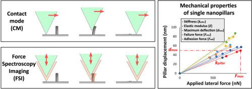
Micro- and nano-patterns are gaining increasing attraction in several fields ranging from nanoelectronics to bioengineering. The mechanical properties of the nanostructures (nanopillars, nanotubes, nanowires, etc.) are highly relevant for many applications but challenging to determine. Existing mechanical characterization methods require mounting the testing setup inside a scanning electron microscope (SEM) and additional sample modification. Here, we propose two atomic force microscopy (AFM) methods, based on contact mode imaging (CMI) and force spectroscopy imaging (FSI), to determine the mechanical characteristics of individual micro- and nanopillars as fabricated, without using SEM. We present the working principles of both methods and two case studies on nanopillars fabricated by additive manufacturing methods: two-photon polymerization (2PP) and electron beam induced deposition (EBID). Various mechanical parameters were determined using CMI and FSI, respectively. For the 2PP nanopillars, we measured the stiffness (13.5 ± 3.2 N/m and 15.9 ± 2.6 N/m), the maximum lateral force (883.0 ± 89.5 nN and 889.6 ± 113.6 nN), the maximum deflection (64.2 ± 13.6 nm and 58.3 ± 14.24 nm), the failure stress (0.3 ± 0.03 GPa and 0.3 ± 0.02 GPa), and the adhesion force (56.6 ± 4.5 µN and 58.6 ± 5.2 µN). For the EBID nanopillars, we measured the failure stress (2.9 ± 0.2 GPa and 2.7 ± 0.4 GPa). The similar results obtained using both techniques confirmed the efficacy and consistency of the methods. The proposed methodologies have the potential of enabling otherwise impossible measurements particularly when the specimens need to be tested under wet conditions, such as patterns for mechanobiological studies.
中文翻译:
原子力显微镜对纳米柱的力学表征
在从纳米电子学到生物工程学的多个领域中,微图案和纳米图案越来越受到人们的关注。纳米结构(纳米柱,纳米管,纳米线等)的机械性能)与许多应用程序高度相关,但很难确定。现有的机械表征方法需要将测试装置安装在扫描电子显微镜(SEM)内,并进行其他样品修改。在这里,我们提出两种基于接触模式成像(CMI)和力谱成像(FSI)的原子力显微镜(AFM)方法,无需使用SEM即可确定所制造的单个微柱和纳米柱的机械特性。我们介绍了两种方法的工作原理,以及通过增材制造方法制造的纳米柱的两个案例研究:双光子聚合(2PP)和电子束诱导沉积(EBID)。分别使用CMI和FSI确定了各种机械参数。对于2PP纳米柱,我们测量了刚度(13.5±3.2 N / m和15.9±2.6 N / m),最大横向力(883.0±89.5 nN和889.6±113.6 nN),最大挠度(64.2±13.6 nm和58.3±14.24 nm),破坏应力(0.3±0.03 GPa和0.3±0.02 GPa)和附着力(56.6±4.5 µN和58.6±5.2 µN)。对于EBID纳米柱,我们测量了破坏应力(2.9±0.2 GPa和2.7±0.4 GPa)。使用这两种技术获得的相似结果证实了该方法的有效性和一致性。所提出的方法具有实现原本不可能进行的测量的潜力,特别是当样品需要在潮湿条件下进行测试时,例如用于力学生物学研究的模式。6±4.5 µN和58.6±5.2 µN)。对于EBID纳米柱,我们测量了破坏应力(2.9±0.2 GPa和2.7±0.4 GPa)。使用这两种技术获得的相似结果证实了该方法的有效性和一致性。所提出的方法具有实现原本不可能进行的测量的潜力,特别是当样品需要在潮湿条件下进行测试时,例如用于力学生物学研究的模式。6±4.5 µN和58.6±5.2 µN)。对于EBID纳米柱,我们测量了破坏应力(2.9±0.2 GPa和2.7±0.4 GPa)。使用这两种技术获得的相似结果证实了该方法的有效性和一致性。所提出的方法具有实现原本不可能进行的测量的潜力,特别是当样品需要在潮湿条件下进行测试时,例如用于力学生物学研究的模式。


 京公网安备 11010802027423号