Our official English website, www.x-mol.net, welcomes your feedback! (Note: you will need to create a separate account there.)
Nanoscale flow cytometry for immunophenotyping and quantitating extracellular vesicles in blood plasma
Nanoscale ( IF 6.7 ) Pub Date : 2021-1-15 , DOI: 10.1039/d0nr05525e Nikki Salmond 1, 2, 3, 4 , Karan Khanna 1, 2, 3, 4 , Gethin R. Owen 2, 5, 6, 7, 8 , Karla C. Williams 1, 2, 3, 4
Nanoscale ( IF 6.7 ) Pub Date : 2021-1-15 , DOI: 10.1039/d0nr05525e Nikki Salmond 1, 2, 3, 4 , Karan Khanna 1, 2, 3, 4 , Gethin R. Owen 2, 5, 6, 7, 8 , Karla C. Williams 1, 2, 3, 4
Affiliation
|
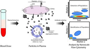
Extracellular vesicles (EVs) are lipid membrane enclosed nano-sized structures released into the extracellular environment by all cell types. EV constituents include proteins, lipids and nucleic acids that reflect the cell from which they originated. The molecular profile of cancer cells is distinct as compared to healthy cells of the same tissue type, and this distinct profile should be reflected by the EVs they release. This makes EVs desirable candidates for blood-based biopsy diagnosis of cancer. EVs can be time consuming to isolate therefore, a technology that can analyze EVs in complex biological samples in a high throughput manner is in demand. Here nanoscale flow cytometry is used to analyze EVs in whole, unpurified, plasma samples from healthy individuals and breast cancer patients. A known breast cancer marker, mammaglobin-a, was evaluated as a potential candidate for expression on EVs and increased levels in breast cancer. Mammaglobin-a particles were abundantly detected in plasma by nanoscale flow cytometry but only a portion of these particles were validated as bona fide EVs. EVs could be distinguish and characterized from small protein clusters and platelets based on size, marker composition, and detergent treatment. Mammaglobin-a positive EVs were characterized and found to be CD42a/CD41-positive platelet EVs, and the number of these EVs present was dependent upon plasma preparation protocol. Different plasma preparation protocols influenced the total number of platelet EVs and mammaglobin-a was found to associate with lipid membranes in plasma. When comparing plasma samples prepared by the same protocol, mammaglobin-a positive EVs were more abundant in estrogen receptor (ER) positive as compared to ER negative breast cancer patient plasma samples. This study demonstrates the capabilities of nanoscale flow cytometry for EV and small particle analysis in whole, unpurified, plasma samples, and highlights important technical challenges that need to be addressed when developing this technology as a liquid biopsy platform.
中文翻译:

纳米级流式细胞仪用于免疫分型和定量血浆中的细胞外囊泡
细胞外囊泡(EVs)是脂质膜包裹的纳米结构,所有类型的细胞都释放到细胞外环境中。电动汽车的成分包括反映其来源细胞的蛋白质,脂质和核酸。与相同组织类型的健康细胞相比,癌细胞的分子特征是截然不同的,并且这种独特的特征应该由它们释放的电动汽车反映出来。这使电动汽车成为进行基于血液的癌症活检诊断的理想候选药物。隔离电动汽车可能很耗时,因此,需要一种能够以高通量方式分析复杂生物样品中电动汽车的技术。在这里,纳米级流式细胞仪用于分析来自健康个体和乳腺癌患者的整个未纯化血浆样品中的电动汽车。已知的乳腺癌标志物,乳房珠蛋白-a,被评估为在电动汽车上表达和增加乳腺癌水平的潜在候选者。通过纳米级流式细胞术在血浆中大量检测到了乳珠蛋白-a颗粒,但这些颗粒中只有一部分被确认为善意电动汽车。电动汽车可根据大小,标记物成分和去污剂处理,与小蛋白簇和血小板区分开并加以表征。哺乳动物珠蛋白a阳性EVs的特征是CD42a / CD41阳性血小板EVs,这些EVs的数量取决于血浆制备方案。不同的血浆制备方案影响了血小板电动车的总数,并且发现血红蛋白-a与血浆中的脂膜有关。当比较通过相同方案制备的血浆样品时,与ER阴性乳腺癌患者血浆样品相比,γ-珠蛋白-a阳性EV的雌激素受体(ER)阳性更为丰富。这项研究证明了纳米级流式细胞仪在整个未纯化血浆样品中进行电动汽车和小颗粒分析的能力,
更新日期:2021-01-15
中文翻译:
纳米级流式细胞仪用于免疫分型和定量血浆中的细胞外囊泡
细胞外囊泡(EVs)是脂质膜包裹的纳米结构,所有类型的细胞都释放到细胞外环境中。电动汽车的成分包括反映其来源细胞的蛋白质,脂质和核酸。与相同组织类型的健康细胞相比,癌细胞的分子特征是截然不同的,并且这种独特的特征应该由它们释放的电动汽车反映出来。这使电动汽车成为进行基于血液的癌症活检诊断的理想候选药物。隔离电动汽车可能很耗时,因此,需要一种能够以高通量方式分析复杂生物样品中电动汽车的技术。在这里,纳米级流式细胞仪用于分析来自健康个体和乳腺癌患者的整个未纯化血浆样品中的电动汽车。已知的乳腺癌标志物,乳房珠蛋白-a,被评估为在电动汽车上表达和增加乳腺癌水平的潜在候选者。通过纳米级流式细胞术在血浆中大量检测到了乳珠蛋白-a颗粒,但这些颗粒中只有一部分被确认为善意电动汽车。电动汽车可根据大小,标记物成分和去污剂处理,与小蛋白簇和血小板区分开并加以表征。哺乳动物珠蛋白a阳性EVs的特征是CD42a / CD41阳性血小板EVs,这些EVs的数量取决于血浆制备方案。不同的血浆制备方案影响了血小板电动车的总数,并且发现血红蛋白-a与血浆中的脂膜有关。当比较通过相同方案制备的血浆样品时,与ER阴性乳腺癌患者血浆样品相比,γ-珠蛋白-a阳性EV的雌激素受体(ER)阳性更为丰富。这项研究证明了纳米级流式细胞仪在整个未纯化血浆样品中进行电动汽车和小颗粒分析的能力,


 京公网安备 11010802027423号