当前位置:
X-MOL 学术
›
Int. J. Dev. Neurosci.
›
论文详情
Our official English website, www.x-mol.net, welcomes your feedback! (Note: you will need to create a separate account there.)
Clinically Detectable Structural Abnormalities in Pediatric Onset Multiple Sclerosis: A Large‐Scale Magnetic Resonance Imaging Analysis
International Journal of Developmental Neuroscience ( IF 1.8 ) Pub Date : 2021-01-29 , DOI: 10.1002/jdn.10090 Jacob Levman 1 , Avilash Das 2 , Allissa MacDonald 3 , Patrick MacDonald 2 , Lindsay Berrigan 4 , Emi Takahashi 2
International Journal of Developmental Neuroscience ( IF 1.8 ) Pub Date : 2021-01-29 , DOI: 10.1002/jdn.10090 Jacob Levman 1 , Avilash Das 2 , Allissa MacDonald 3 , Patrick MacDonald 2 , Lindsay Berrigan 4 , Emi Takahashi 2
Affiliation
|
BACKGROUND
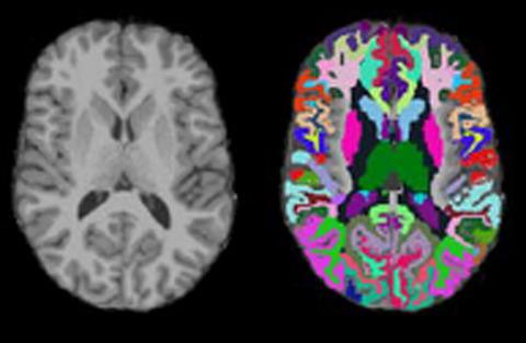
Multiple Sclerosis is characterized by neural demyelination. Structural magnetic resonance imaging (MRI) provides soft tissue contrast, which forms the basis of techniques for extracting regional biomarkers across a participant's brain. OBJECTIVES
To investigate the clinical presentation of multiple sclerosis in a large-scale MRI analysis that includes thorough consideration of extractable structural measurements (average and variability of regional cortical thicknesses, cortical surface measurements and volumes). METHODS
We performed a large-scale retrospective analysis of 370 T1 structural volumetric MRIs from 64 participants with multiple sclerosis and compared them with a large cohort of neurotypical participants, consisting of 993 MRIs from 988 participants. Regionally distributed measurements of cortical thickness (average and standard deviation) were extracted along with surface area, surface curvature and volumetric measurements. RESULTS
The largest observed finding involved regionally distributed reductions in average cortical thickness, with the parahippocampal region exhibiting the largest effect size, a finding that may be linked with known hippocampal atrophy in multiple sclerosis. Group-wise differences were also observed in terms of distributed volume, surface area and surface curvature measurements. CONCLUSIONS
Participants with pediatric onset multiple sclerosis present clinically with a variety of structural abnormalities, including perirhinal cortex thickness abnormalities not previously reported in the literature.
中文翻译:

小儿多发性硬化症的临床可检测结构异常:大规模磁共振成像分析
背景多发性硬化的特征在于神经脱髓鞘。结构磁共振成像 (MRI) 提供软组织对比度,它构成了在参与者大脑中提取区域生物标志物的技术基础。目的 在大规模 MRI 分析中调查多发性硬化症的临床表现,包括全面考虑可提取的结构测量值(区域皮质厚度、皮质表面测量值和体积的平均值和变异性)。方法我们对来自 64 名多发性硬化症参与者的 370 次 T1 结构体积 MRI 进行了大规模回顾性分析,并将它们与一大群神经典型参与者进行了比较,该队列由来自 988 名参与者的 993 次 MRI 组成。皮层厚度(平均值和标准偏差)的区域分布测量值与表面积、表面曲率和体积测量值一起被提取。结果 观察到的最大发现涉及平均皮质厚度的区域分布减少,海马旁区域表现出最大的影响大小,这一发现可能与多发性硬化症中已知的海马萎缩有关。在分布体积、表面积和表面曲率测量方面也观察到组间差异。结论 患有小儿多发性硬化症的参与者在临床上表现出各种结构异常,包括以前文献中未报道的周围皮层厚度异常。表面曲率和体积测量。结果 观察到的最大发现涉及平均皮质厚度的区域分布减少,海马旁区域表现出最大的影响大小,这一发现可能与多发性硬化症中已知的海马萎缩有关。在分布体积、表面积和表面曲率测量方面也观察到组间差异。结论 患有小儿多发性硬化症的参与者在临床上表现出各种结构异常,包括以前文献中未报道的周围皮层厚度异常。表面曲率和体积测量。结果 观察到的最大发现涉及平均皮质厚度的区域分布减少,海马旁区域表现出最大的影响大小,这一发现可能与多发性硬化症中已知的海马萎缩有关。在分布体积、表面积和表面曲率测量方面也观察到组间差异。结论 患有小儿多发性硬化症的参与者在临床上表现出各种结构异常,包括以前文献中未报道的周围皮层厚度异常。海马旁区域表现出最大的效应量,这一发现可能与多发性硬化症中已知的海马萎缩有关。在分布体积、表面积和表面曲率测量方面也观察到组间差异。结论 患有小儿多发性硬化症的参与者在临床上表现出各种结构异常,包括以前文献中未报道的周围皮层厚度异常。海马旁区域表现出最大的效应量,这一发现可能与多发性硬化症中已知的海马萎缩有关。在分布体积、表面积和表面曲率测量方面也观察到组间差异。结论 患有小儿多发性硬化症的参与者在临床上表现出各种结构异常,包括以前文献中未报道的周围皮层厚度异常。
更新日期:2021-01-29
中文翻译:
小儿多发性硬化症的临床可检测结构异常:大规模磁共振成像分析
背景多发性硬化的特征在于神经脱髓鞘。结构磁共振成像 (MRI) 提供软组织对比度,它构成了在参与者大脑中提取区域生物标志物的技术基础。目的 在大规模 MRI 分析中调查多发性硬化症的临床表现,包括全面考虑可提取的结构测量值(区域皮质厚度、皮质表面测量值和体积的平均值和变异性)。方法我们对来自 64 名多发性硬化症参与者的 370 次 T1 结构体积 MRI 进行了大规模回顾性分析,并将它们与一大群神经典型参与者进行了比较,该队列由来自 988 名参与者的 993 次 MRI 组成。皮层厚度(平均值和标准偏差)的区域分布测量值与表面积、表面曲率和体积测量值一起被提取。结果 观察到的最大发现涉及平均皮质厚度的区域分布减少,海马旁区域表现出最大的影响大小,这一发现可能与多发性硬化症中已知的海马萎缩有关。在分布体积、表面积和表面曲率测量方面也观察到组间差异。结论 患有小儿多发性硬化症的参与者在临床上表现出各种结构异常,包括以前文献中未报道的周围皮层厚度异常。表面曲率和体积测量。结果 观察到的最大发现涉及平均皮质厚度的区域分布减少,海马旁区域表现出最大的影响大小,这一发现可能与多发性硬化症中已知的海马萎缩有关。在分布体积、表面积和表面曲率测量方面也观察到组间差异。结论 患有小儿多发性硬化症的参与者在临床上表现出各种结构异常,包括以前文献中未报道的周围皮层厚度异常。表面曲率和体积测量。结果 观察到的最大发现涉及平均皮质厚度的区域分布减少,海马旁区域表现出最大的影响大小,这一发现可能与多发性硬化症中已知的海马萎缩有关。在分布体积、表面积和表面曲率测量方面也观察到组间差异。结论 患有小儿多发性硬化症的参与者在临床上表现出各种结构异常,包括以前文献中未报道的周围皮层厚度异常。海马旁区域表现出最大的效应量,这一发现可能与多发性硬化症中已知的海马萎缩有关。在分布体积、表面积和表面曲率测量方面也观察到组间差异。结论 患有小儿多发性硬化症的参与者在临床上表现出各种结构异常,包括以前文献中未报道的周围皮层厚度异常。海马旁区域表现出最大的效应量,这一发现可能与多发性硬化症中已知的海马萎缩有关。在分布体积、表面积和表面曲率测量方面也观察到组间差异。结论 患有小儿多发性硬化症的参与者在临床上表现出各种结构异常,包括以前文献中未报道的周围皮层厚度异常。


 京公网安备 11010802027423号