Computers in Biology and Medicine ( IF 7.7 ) Pub Date : 2021-01-11 , DOI: 10.1016/j.compbiomed.2021.104214 Fernando O Campos 1 , Michele Orini 2 , Robert Arnold 3 , John Whitaker 1 , Mark O'Neill 1 , Reza Razavi 1 , Gernot Plank 3 , Ben Hanson 4 , Bradley Porter 5 , Christopher Aldo Rinaldi 6 , Jaswinder Gill 5 , Pier D Lambiase 2 , Peter Taggart 2 , Martin J Bishop 1
|
Background
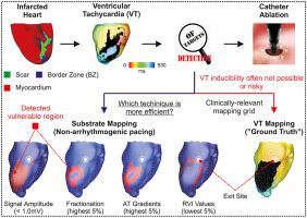
Identification of targets for ablation of post-infarction ventricular tachycardias (VTs) remains challenging, often requiring arrhythmia induction to delineate the reentrant circuit. This carries a risk for the patient and may not be feasible. Substrate mapping has emerged as a safer strategy to uncover arrhythmogenic regions. However, VT recurrence remains common.
Goal
To use computer simulations to assess the ability of different substrate mapping approaches to identify VT exit sites.
Methods
A 3D computational model of the porcine post-infarction heart was constructed to simulate VT and paced rhythm. Electroanatomical maps were constructed based on endocardial electrogram features and the reentry vulnerability index (RVI - a metric combining activation (AT) and repolarization timings to identify tissue susceptibility to reentry). Since scar transmurality in our model was not homogeneous, parameters derived from all signals (including dense scar regions) were used in the analysis. Potential ablation targets obtained from each electroanatomical map during pacing were compared to the exit site detected during VT mapping.
Results
Simulation data showed that voltage cut-offs applied to bipolar electrograms could delineate the scar, but not the VT circuit. Electrogram fractionation had the highest correlation with scar transmurality. The RVI identified regions closest to VT exit site but was outperformed by AT gradients combined with voltage cut-offs. The performance of all metrics was affected by pacing location.
Conclusions
Substrate mapping could provide information about the infarct, but the directional dependency on activation should be considered. Activation-repolarization metrics have utility in safely identifying VT targets, even with non-transmural scars.
中文翻译:
使用计算模型评估底物映射技术指导室性心动过速消融的能力
背景
确定用于消融梗死后室性心动过速 (VT) 的目标仍然具有挑战性,通常需要心律失常诱导来描绘折返回路。这会给患者带来风险并且可能不可行。底物映射已成为发现致心律失常区域的一种更安全的策略。然而,VT 复发仍然很常见。
目标
使用计算机模拟来评估不同底物映射方法识别 VT 出口位置的能力。
方法
构建了猪梗死后心脏的 3D 计算模型以模拟 VT 和起搏节律。电解剖图是根据心内膜电图特征和折返脆弱性指数(RVI - 一种结合激活 (AT) 和复极时间来识别组织折返易感性的指标)构建的。由于我们模型中的疤痕透壁性不均匀,因此在分析中使用了来自所有信号(包括密集疤痕区域)的参数。将起搏期间从每个电解剖图获得的潜在消融目标与 VT 标测期间检测到的出口部位进行比较。
结果
模拟数据显示,应用于双极电图的电压截止可以描绘疤痕,但不能描绘 VT 电路。电图分割与疤痕透壁性的相关性最高。RVI 确定了最接近 VT 出口位置的区域,但 AT 梯度与电压截止相结合的表现优于该区域。所有指标的表现都受到起搏位置的影响。
结论
底物映射可以提供有关梗塞的信息,但应考虑对激活的方向依赖性。激活-复极指标可用于安全识别 VT 目标,即使是非透壁疤痕。


 京公网安备 11010802027423号