Medical & Biological Engineering & Computing ( IF 3.2 ) Pub Date : 2021-01-07 , DOI: 10.1007/s11517-020-02302-w Panagiotis Marentakis 1 , Pantelis Karaiskos 1 , Vassilis Kouloulias 2 , Nikolaos Kelekis 2 , Stylianos Argentos 2 , Nikolaos Oikonomopoulos 2 , Constantinos Loukas 1
|
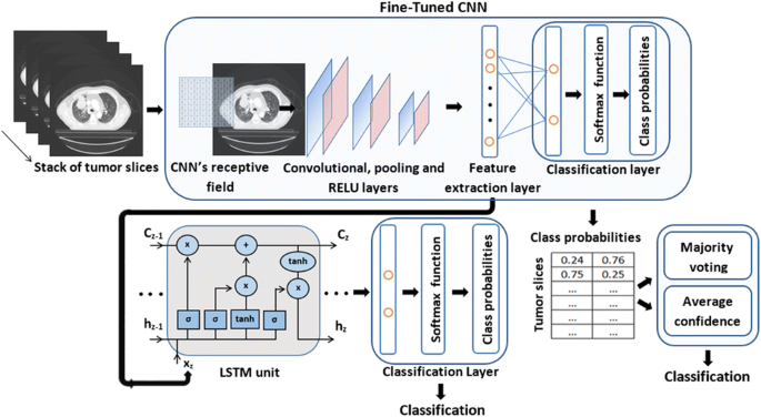
Adenocarcinoma (AC) and squamous cell carcinoma (SCC) are frequent reported cases of non-small cell lung cancer (NSCLC), responsible for a large fraction of cancer deaths worldwide. In this study, we aim to investigate the potential of NSCLC histology classification into AC and SCC by applying different feature extraction and classification techniques on pre-treatment CT images. The employed image dataset (102 patients) was taken from the publicly available cancer imaging archive collection (TCIA). We investigated four different families of techniques: (a) radiomics with two classifiers (kNN and SVM), (b) four state-of-the-art convolutional neural networks (CNNs) with transfer learning and fine tuning (Alexnet, ResNet101, Inceptionv3 and InceptionResnetv2), (c) a CNN combined with a long short-term memory (LSTM) network to fuse information about the spatial coherency of tumor’s CT slices, and (d) combinatorial models (LSTM + CNN + radiomics). In addition, the CT images were independently evaluated by two expert radiologists. Our results showed that the best CNN was Inception (accuracy = 0.67, auc = 0.74). LSTM + Inception yielded superior performance than all other methods (accuracy = 0.74, auc = 0.78). Moreover, LSTM + Inception outperformed experts by 7–25% (p < 0.05). The proposed methodology does not require detailed segmentation of the tumor region and it may be used in conjunction with radiological findings to improve clinical decision-making.
Graphical abstract
中文翻译:
基于影像组学和深度学习模型的 CT 图像肺癌组织学分类
腺癌 (AC) 和鳞状细胞癌 (SCC) 是非小细胞肺癌 (NSCLC) 的常见报告病例,它们是全球癌症死亡的很大一部分。在本研究中,我们旨在通过对治疗前 CT 图像应用不同的特征提取和分类技术来研究 NSCLC 组织学分类为 AC 和 SCC 的潜力。所采用的图像数据集(102 名患者)取自公开可用的癌症成像档案馆 (TCIA)。我们研究了四种不同的技术系列:(a)具有两个分类器(kNN 和 SVM)的放射组学,(b)具有迁移学习和微调的四个最先进的卷积神经网络(CNN)(Alexnet、ResNet101、Inceptionv3和 InceptionResnetv2), (c) CNN 与长短期记忆 (LSTM) 网络相结合,以融合有关肿瘤 CT 切片空间一致性的信息,以及 (d) 组合模型(LSTM + CNN + 放射组学)。此外,CT 图像由两名放射科专家独立评估。我们的结果表明,最好的 CNN 是 Inception(准确率 = 0.67,auc = 0.74)。LSTM + Inception 产生了优于所有其他方法的性能(准确度 = 0.74,auc = 0.78)。此外,LSTM + Inception 的表现比专家高 7-25%(74,auc = 0.78)。此外,LSTM + Inception 的表现比专家高 7-25%(74,auc = 0.78)。此外,LSTM + Inception 的表现比专家高 7-25%(p < 0.05)。所提出的方法不需要对肿瘤区域进行详细分割,并且可以与放射学发现结合使用以改进临床决策。


 京公网安备 11010802027423号