Biomaterials Advances ( IF 7.9 ) Pub Date : 2021-01-06 , DOI: 10.1016/j.msec.2020.111858 Yuqing Niu 1 , Florian J Stadler 2 , Ming Fu 1
|
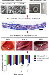
The micro- or nanoscale surface morphology of the tissue engineering nerve guidance scaffold (NGS) will affect different cell behaviors, such as their growth rate, migration, and matrix secretion. Although different technologies for manufacturing scaffolds with biomimetic topography have been established, most of them tend to be high cost and long preparation time. Here we have prepared a biomimetic NGS with physical properties to simulate native nerve tissue more accurately. We used poly(l-lactic acid) (PLLA) nanofibers doped with gelatin to prepare a biomimetic NGS whose structure mimics the native epineurium layer. By adjusting the doping ratio of gelatin and PLLA in the tubular scaffold, the bionic scaffold's surface morphology and mechanical properties are closer to native tissues. In vitro cell scaffold interaction experiments demonstrated that the PLLA/gelatin nanofibers could significantly promote the elongation, proliferation, and the secretion of glial cell-derived neurotrophic factor (GDNF) of RSC96 Schwann cells (SCs), as well as the diffusion of GDNF. In vivo scaffold replacement of SD rat, sciatic nerves showed that the nerve guide scaffold composed of PLLA/gelatin nanofibers was helpful to the myelination of SCs and the remolding of epineurium in the injured area, which could effectively rehabilitate the motor and sensory functions of the injured nerve and prevent the atrophy of the target muscle tissue. This study showed that the synergistic impact of nano topographical and biochemical clues on designing biomimetic scaffolds could efficiently promote regenerating nerve tissue.
中文翻译:
仿生电纺管状 PLLA/明胶纳米纤维支架促进 SD 大鼠坐骨神经断面再生
组织工程神经引导支架(NGS)的微米或纳米级表面形态将影响不同的细胞行为,例如它们的生长速率、迁移和基质分泌。尽管已经建立了不同的用于制造具有仿生形貌的支架的技术,但大多数技术往往成本高且准备时间长。在这里,我们制备了具有物理特性的仿生NGS,可以更准确地模拟天然神经组织。我们使用掺杂明胶的聚(L-乳酸)(PLLA)纳米纤维来制备仿生NGS,其结构模仿天然神经外膜层。通过调整管状支架中明胶和PLLA的掺杂比例,仿生支架的表面形貌和力学性能更接近天然组织。体外细胞支架相互作用实验表明,PLLA/明胶纳米纤维可以显着促进RSC96雪旺细胞(SC)的伸长、增殖、胶质细胞源性神经营养因子(GDNF)的分泌以及GDNF的扩散。SD大鼠坐骨神经体内支架置换结果表明,PLLA/明胶纳米纤维组成的神经导向支架有助于损伤部位SCs的髓鞘化和神经外膜的重塑,可有效恢复其运动和感觉功能。损伤神经并防止目标肌肉组织萎缩。这项研究表明,纳米拓扑和生化线索在设计仿生支架时的协同作用可以有效促进神经组织的再生。


 京公网安备 11010802027423号