Journal of Neuroradiology ( IF 3.5 ) Pub Date : 2021-01-05 , DOI: 10.1016/j.neurad.2020.12.004 Rupa Radhakrishnan 1 , Brandon P Brown 1 , David M Haas 2 , Yong Zang 3 , Christina Sparks 4 , Senthilkumar Sadhasivam 4
|
Purpose
The purpose of this study was to assess for any differences in brain maturation, structure and morphometry in fetuses exposed to opioids in utero, compared to non-opioid exposed fetuses on fetal MRI.
Methods
We performed a prospective study in pregnant women using opioids and healthy pregnant women without prenatal opioid use. We evaluated brain maturation, structure, and morphometry on second or third trimester fetal MRI and assessed group differences.
Results
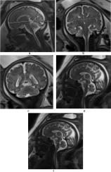
28 pregnant women were enrolled, 12 with opioid exposure (average gestational age 33.67, range 28–39 w), 9 of whom also smoked, and 16 without opioid exposure (average gestational age 32.53, range 27–38 w). There was a significant difference in the anteroposterior diameter of the fetal cerebellar vermis in the opioid exposed fetuses compared to non-opioid exposed fetuses (p = 0.004). There were no significant differences in brain biparietal diameter, fronto-occipital diameter, transverse cerebellar diameter and anteroposterior dimension of the pons in opioid exposed fetuses compared to non-opioid exposed fetuses. There were no abnormalities in brain maturation and no major brain structural abnormalities in the opioid exposed fetuses.
Conclusion
Smaller fetal anteroposterior cerebellar vermian dimension was associated with in utero opioid exposure. There were no abnormalities in brain maturation or major structural abnormalities in fetuses exposed to opioids.
中文翻译:
胎儿大脑发育和形态测定在胎儿 MRI 上产前阿片类药物暴露和吸烟的初步研究
目的
本研究的目的是通过胎儿 MRI 评估与未接触阿片类药物的胎儿相比,在子宫内接触阿片类药物的胎儿在大脑成熟、结构和形态测量方面的任何差异。
方法
我们对使用阿片类药物的孕妇和产前未使用阿片类药物的健康孕妇进行了一项前瞻性研究。我们评估了妊娠中期或晚期胎儿 MRI 的大脑成熟度、结构和形态计量学,并评估了组间差异。
结果
招募了 28 名孕妇,其中 12 名暴露于阿片类药物(平均胎龄 33.67,范围 28-39 周),其中 9 名还吸烟,16 名未暴露于阿片类药物(平均胎龄 32.53,范围 27-38 周)。与非阿片类药物暴露胎儿相比,阿片类药物暴露胎儿的胎儿小脑蚓部前后径存在显着差异 (p = 0.004)。与非阿片类药物暴露胎儿相比,阿片类药物暴露胎儿的脑双顶径、额枕直径、小脑横径和脑桥前后径无显着差异。暴露于阿片类药物的胎儿的大脑成熟没有异常,也没有重大的大脑结构异常。
结论
较小的胎儿前后小脑蚓部尺寸与子宫内阿片类药物暴露有关。暴露于阿片类药物的胎儿没有大脑成熟异常或主要结构异常。


 京公网安备 11010802027423号