当前位置:
X-MOL 学术
›
Hum. Brain Mapp.
›
论文详情
Our official English website, www.x-mol.net, welcomes your feedback! (Note: you will need to create a separate account there.)
Decreasing brain iron in multiple sclerosis: The difference between concentration and content in iron MRI
Human Brain Mapping ( IF 4.8 ) Pub Date : 2020-12-30 , DOI: 10.1002/hbm.25306 Ferdinand Schweser 1, 2 , Jesper Hagemeier 1 , Michael G Dwyer 1, 2 , Niels Bergsland 1, 3 , Simon Hametner 4 , Bianca Weinstock-Guttman 5 , Robert Zivadinov 1, 2
Human Brain Mapping ( IF 4.8 ) Pub Date : 2020-12-30 , DOI: 10.1002/hbm.25306 Ferdinand Schweser 1, 2 , Jesper Hagemeier 1 , Michael G Dwyer 1, 2 , Niels Bergsland 1, 3 , Simon Hametner 4 , Bianca Weinstock-Guttman 5 , Robert Zivadinov 1, 2
Affiliation
|
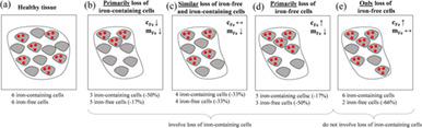
Increased brain iron concentration is often reported concurrently with disease development in multiple sclerosis (MS) and other neurodegenerative diseases. However, it is unclear whether the higher iron concentration in patients stems from an influx of iron into the tissue or a relative reduction in tissue compartments without much iron. By taking into account structural volume, we investigated tissue iron content in the deep gray matter (DGM) over 2 years, and compared findings to previously reported changes in iron concentration. 120 MS patients and 40 age‐ and sex‐matched healthy controls were included. Clinical testing and MRI were performed both at baseline and after 2 years. Overall, iron content was calculated from structural MRI and quantitative susceptibility mapping in the thalamus, caudate, putamen, and globus pallidus. MS patients had significantly lower iron content than controls in the thalamus, with progressive MS patients demonstrating lower iron content than relapsing–remitting patients. Over 2 years, iron content decreased in the DGM of patients with MS, while it tended to increase or remain stable among controls. In the thalamus, decreasing iron content over 2 years was associated with disability progression. Our study showed that temporally increasing magnetic susceptibility in MS should not be considered as evidence for iron influx because it may be explained, at least partially, by disease‐related atrophy. Declining DGM iron content suggests that, contrary to the current understanding, iron is being removed from the DGM in patients with MS.
中文翻译:

减少多发性硬化症中的脑铁:铁 MRI 中浓度和含量的差异
在多发性硬化症 (MS) 和其他神经退行性疾病的疾病发展过程中,经常报告脑铁浓度增加。然而,目前尚不清楚患者体内较高的铁浓度是由于铁流入组织还是由于铁含量不足的组织隔室相对减少。通过考虑结构体积,我们在 2 年内调查了深部灰质 (DGM) 中的组织铁含量,并将结果与之前报道的铁浓度变化进行了比较。包括 120 名 MS 患者和 40 名年龄和性别匹配的健康对照。在基线和 2 年后进行了临床测试和 MRI。综合铁含量由丘脑、尾状核、壳核和苍白球的结构 MRI 和定量磁化率映射计算。MS 患者的丘脑铁含量显着低于对照组,进展性 MS 患者的铁含量低于复发缓解患者。2 年多来,MS 患者 DGM 中的铁含量下降,而对照组中铁含量趋于增加或保持稳定。在丘脑中,2 年内铁含量下降与残疾进展相关。我们的研究表明,MS 中暂时增加的磁化率不应被视为铁内流的证据,因为它至少可以部分地由疾病相关的萎缩来解释。DGM铁含量下降 表明,与目前的理解相反,正在从 MS 患者的 DGM 中去除铁。
更新日期:2021-03-03
中文翻译:
减少多发性硬化症中的脑铁:铁 MRI 中浓度和含量的差异
在多发性硬化症 (MS) 和其他神经退行性疾病的疾病发展过程中,经常报告脑铁浓度增加。然而,目前尚不清楚患者体内较高的铁浓度是由于铁流入组织还是由于铁含量不足的组织隔室相对减少。通过考虑结构体积,我们在 2 年内调查了深部灰质 (DGM) 中的组织铁含量,并将结果与之前报道的铁浓度变化进行了比较。包括 120 名 MS 患者和 40 名年龄和性别匹配的健康对照。在基线和 2 年后进行了临床测试和 MRI。综合铁含量由丘脑、尾状核、壳核和苍白球的结构 MRI 和定量磁化率映射计算。MS 患者的丘脑铁含量显着低于对照组,进展性 MS 患者的铁含量低于复发缓解患者。2 年多来,MS 患者 DGM 中的铁含量下降,而对照组中铁含量趋于增加或保持稳定。在丘脑中,2 年内铁含量下降与残疾进展相关。我们的研究表明,MS 中暂时增加的磁化率不应被视为铁内流的证据,因为它至少可以部分地由疾病相关的萎缩来解释。DGM铁含量下降 表明,与目前的理解相反,正在从 MS 患者的 DGM 中去除铁。


 京公网安备 11010802027423号