Biochimica et Biophysica Acta (BBA) - Bioenergetics ( IF 4.3 ) Pub Date : 2020-12-29 , DOI: 10.1016/j.bbabio.2020.148366 Leyla Rohani 1 , Gary Hastings 2
|
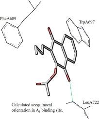
Time resolved FTIR difference spectroscopy (DS) has been used to study photosystem I (PSI) with the disubstituted 1,4-naphthoquinones acequinocyl (AcQ) and lapachol (Lpc) incorporated into the A1 binding site. AcQ is a 2-acetoxy-3-dodecyl-1,4-naphthoquinone, Lpc is a 2-hydroxy-3-(3-methyl-2-butenyl)-1,4-naphthoquinone. To assess whether the experimental spectra are specific to different orientations of the quinone and their substitutions ONIOM-type QM/MM vibrational frequency calculations were undertaken for various orientations of the pigments and side-chain conformations in the A1 binding site.
Comparison of calculated and experimental spectra for the reduced species (semiquinone anion) suggests that the orientation for the naphthoquinone ring in the binding site and specific side-chain conformations can be identified based on the spectra. In native PSI phylloquinone (PhQ) in the A1 binding site binds with its phytyl chain ortho to the hydrogen bonded carbonyl group. This is not found to be the case for the hydrocarbon tail of AcQ, which is meta to the H-bonded carbonyl group. In contrast, Lpc in PSI binds with its hydrocarbon tail also ortho to the H-bonded carbonyl group. Furthermore, comparison of calculated and experimental spectra indicates which conformations the acetoxy group of AcQ and the hydroxy group of Lpc adopt in the A1 binding site.
中文翻译:
从红外差异光谱评估蛋白质结合位点中色素的取向和构象
时间分辨FTIR差异光谱法(DS)已用于研究光系统I(PSI),并将双取代的1,4-萘醌乙酰喹啉(AcQ)和拉帕胆(Lpc)纳入A 1结合位点。AcQ是2-乙酰氧基-3-十二烷基-1,4-萘醌,Lpc是2-羟基-3-(3-甲基-2-丁烯基)-1,4-萘醌。为了评估实验光谱是否特定于醌的不同方向及其取代,对颜料的各种方向和A 1结合位点的侧链构象进行了ONIOM型QM / MM振动频率计算。
还原物种(半醌阴离子)的计算光谱和实验光谱的比较表明,可以根据光谱识别结合位点中萘醌环的取向和特定的侧链构象。在天然的PSI中,叶绿醌(PhQ)在A 1的结合位点的植酸基链与氢键结合的羰基邻位结合。对于AcQ的烃尾(对于H键合的羰基是间位的),情况并非如此。相反,PSI中的Lpc与其烃尾结合,也与H键合的羰基邻位结合。此外,计算和实验光谱的比较表明,AcQ的乙酰氧基和Lpc的羟基在A 1结合位点采用哪种构象。


 京公网安备 11010802027423号