当前位置:
X-MOL 学术
›
Microsc. Res. Tech.
›
论文详情
Our official English website, www.x-mol.net, welcomes your feedback! (Note: you will need to create a separate account there.)
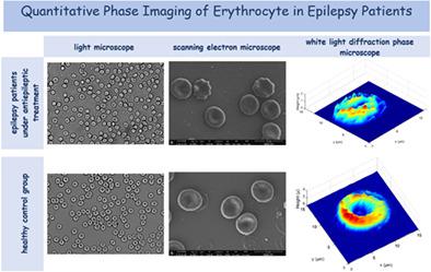
Quantitative phase imaging of erythrocyte in epilepsy patients
Microscopy Research and Technique ( IF 2.5 ) Pub Date : 2020-12-19 , DOI: 10.1002/jemt.23676 Aysun Ünal 1 , Özlem Kocahan 2 , Bengü Altunan 1 , Aslı Aksoy Gündoğdu 1 , Merve Uyanık 2 , Serhat Özder 3
Microscopy Research and Technique ( IF 2.5 ) Pub Date : 2020-12-19 , DOI: 10.1002/jemt.23676 Aysun Ünal 1 , Özlem Kocahan 2 , Bengü Altunan 1 , Aslı Aksoy Gündoğdu 1 , Merve Uyanık 2 , Serhat Özder 3
Affiliation
|
The present study focuses on the quantitative phase imaging of erythrocytes with the aim to compare the morphological differences between epilepsy patients under antiepileptic treatment, who have no other disease which may affect the erythrocyte morphology, and the healthy control group. The white light diffraction phase microscopy (WDPM) has been used to obtain the interferogram of the erythrocyte surfaces. The continuous wavelet transform with Paul wavelet has been chosen to calculate the surface profiles from this interferogram image. For the determination of alteration in morphology, besides WDPM, erythrocyte surfaces have been investigated by light microscope and scanning electron microscope. In this way, it has been possible to see the difference in terms of precision and implementation between the most commonly used methods with regard to the quantitative phase imaging. Erythrocytes from all the samples have been examined and displayed in both two- and three-dimensional way. We have observed that erythrocytes of patients with effective antiepileptic blood levels were more affected in morphology than healthy subjects. When we compared the erythrocyte morphological changes of patients who received monotherapy or polytherapy, no difference was observed. In conclusion, antiepileptic drugs (AEDs) cause red blood cell (RBC) morphological changes and a combined usage of WDPM with Paul wavelet and light microscopy methods are very convenient for studying the erythrocyte morphologies on multiple patients.
中文翻译:

癫痫患者红细胞的定量相位成像
本研究侧重于红细胞的定量相位成像,目的是比较抗癫痫治疗下没有其他可能影响红细胞形态的疾病的癫痫患者与健康对照组之间的形态学差异。白光衍射相位显微镜 (WDPM) 已被用于获得红细胞表面的干涉图。已选择使用 Paul 小波的连续小波变换来计算此干涉图图像的表面轮廓。对于形态学改变的测定,除了 WDPM 外,还通过光学显微镜和扫描电子显微镜研究了红细胞表面。这样,在定量相位成像方面,最常用的方法之间在精度和实施方面的差异是可能的。来自所有样品的红细胞已被检查并以二维和三维方式显示。我们观察到,与健康受试者相比,具有有效抗癫痫血药水平的患者的红细胞在形态上受到的影响更大。当我们比较接受单药治疗或多药治疗的患者的红细胞形态变化时,没有观察到差异。总之,抗癫痫药物(AEDs)会引起红细胞(RBC)形态变化,WDPM与保罗小波和光学显微镜方法的结合使用非常方便研究多个患者的红细胞形态。来自所有样品的红细胞已被检查并以二维和三维方式显示。我们观察到,与健康受试者相比,具有有效抗癫痫血药水平的患者的红细胞在形态上受到的影响更大。当我们比较接受单药治疗或多药治疗的患者的红细胞形态变化时,没有观察到差异。总之,抗癫痫药物(AEDs)会引起红细胞(RBC)形态变化,WDPM与保罗小波和光学显微镜方法的结合使用非常方便研究多个患者的红细胞形态。来自所有样本的红细胞都已被检查并以二维和三维方式显示。我们观察到,与健康受试者相比,具有有效抗癫痫血药水平的患者的红细胞在形态上受到的影响更大。当我们比较接受单药治疗或多药治疗的患者的红细胞形态变化时,没有观察到差异。总之,抗癫痫药物(AEDs)会引起红细胞(RBC)形态变化,WDPM与保罗小波和光学显微镜方法的结合使用非常方便研究多个患者的红细胞形态。我们观察到,与健康受试者相比,具有有效抗癫痫血药水平的患者的红细胞在形态上受到的影响更大。当我们比较接受单药治疗或多药治疗的患者的红细胞形态变化时,没有观察到差异。总之,抗癫痫药物(AEDs)会引起红细胞(RBC)形态变化,WDPM与保罗小波和光学显微镜方法的结合使用非常方便研究多个患者的红细胞形态。我们观察到,与健康受试者相比,具有有效抗癫痫血药水平的患者的红细胞在形态上受到的影响更大。当我们比较接受单药治疗或多药治疗的患者的红细胞形态变化时,没有观察到差异。总之,抗癫痫药物(AEDs)会引起红细胞(RBC)形态变化,WDPM与保罗小波和光学显微镜方法的结合使用非常方便研究多个患者的红细胞形态。
更新日期:2020-12-19
中文翻译:
癫痫患者红细胞的定量相位成像
本研究侧重于红细胞的定量相位成像,目的是比较抗癫痫治疗下没有其他可能影响红细胞形态的疾病的癫痫患者与健康对照组之间的形态学差异。白光衍射相位显微镜 (WDPM) 已被用于获得红细胞表面的干涉图。已选择使用 Paul 小波的连续小波变换来计算此干涉图图像的表面轮廓。对于形态学改变的测定,除了 WDPM 外,还通过光学显微镜和扫描电子显微镜研究了红细胞表面。这样,在定量相位成像方面,最常用的方法之间在精度和实施方面的差异是可能的。来自所有样品的红细胞已被检查并以二维和三维方式显示。我们观察到,与健康受试者相比,具有有效抗癫痫血药水平的患者的红细胞在形态上受到的影响更大。当我们比较接受单药治疗或多药治疗的患者的红细胞形态变化时,没有观察到差异。总之,抗癫痫药物(AEDs)会引起红细胞(RBC)形态变化,WDPM与保罗小波和光学显微镜方法的结合使用非常方便研究多个患者的红细胞形态。来自所有样品的红细胞已被检查并以二维和三维方式显示。我们观察到,与健康受试者相比,具有有效抗癫痫血药水平的患者的红细胞在形态上受到的影响更大。当我们比较接受单药治疗或多药治疗的患者的红细胞形态变化时,没有观察到差异。总之,抗癫痫药物(AEDs)会引起红细胞(RBC)形态变化,WDPM与保罗小波和光学显微镜方法的结合使用非常方便研究多个患者的红细胞形态。来自所有样本的红细胞都已被检查并以二维和三维方式显示。我们观察到,与健康受试者相比,具有有效抗癫痫血药水平的患者的红细胞在形态上受到的影响更大。当我们比较接受单药治疗或多药治疗的患者的红细胞形态变化时,没有观察到差异。总之,抗癫痫药物(AEDs)会引起红细胞(RBC)形态变化,WDPM与保罗小波和光学显微镜方法的结合使用非常方便研究多个患者的红细胞形态。我们观察到,与健康受试者相比,具有有效抗癫痫血药水平的患者的红细胞在形态上受到的影响更大。当我们比较接受单药治疗或多药治疗的患者的红细胞形态变化时,没有观察到差异。总之,抗癫痫药物(AEDs)会引起红细胞(RBC)形态变化,WDPM与保罗小波和光学显微镜方法的结合使用非常方便研究多个患者的红细胞形态。我们观察到,与健康受试者相比,具有有效抗癫痫血药水平的患者的红细胞在形态上受到的影响更大。当我们比较接受单药治疗或多药治疗的患者的红细胞形态变化时,没有观察到差异。总之,抗癫痫药物(AEDs)会引起红细胞(RBC)形态变化,WDPM与保罗小波和光学显微镜方法的结合使用非常方便研究多个患者的红细胞形态。


 京公网安备 11010802027423号