当前位置:
X-MOL 学术
›
Cell. Microbiol.
›
论文详情
Our official English website, www.x-mol.net, welcomes your feedback! (Note: you will need to create a separate account there.)
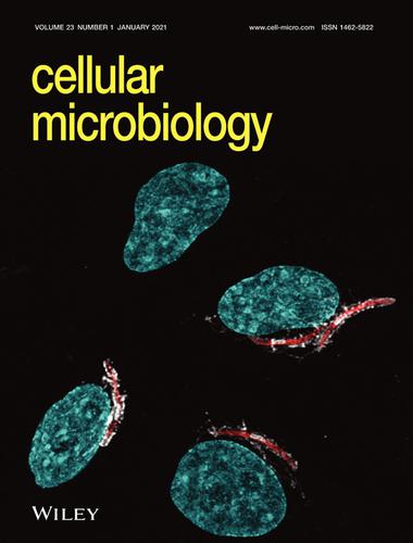
Cover Image: Plasmodium berghei sporozoites in nonreplicative vacuole are eliminated by a PI3P‐mediated autophagy‐independent pathway (Cellular Microbiology 1/2021)
Cellular Microbiology ( IF 3.4 ) Pub Date : 2020-12-07 , DOI: 10.1111/cmi.13292 Annina Bindschedler , Rahel Wacker , Jessica Egli , Nina Eickel , Jacqueline Schmuckli‐Maurer , Blandine M. Franke‐Fayard , Chris J. Janse , Volker T. Heussler
Cellular Microbiology ( IF 3.4 ) Pub Date : 2020-12-07 , DOI: 10.1111/cmi.13292 Annina Bindschedler , Rahel Wacker , Jessica Egli , Nina Eickel , Jacqueline Schmuckli‐Maurer , Blandine M. Franke‐Fayard , Chris J. Janse , Volker T. Heussler
|
Immunofluorescence image of Plasmodium berghei sporozoites (red) in HeLa cells 1.5 hours post infection. The parasitophorous vacuole membrane (PVM) that surrounds the parasites was stained with antibodies against the PVM‐resident protein UIS4 (grey). DNA was stained with DAPI (blue). Imaging was performed on a Leica SP8 confocal microscope. For further details, readers are referred to the article by Bindschedler et al. on p. e13271 of this issue.

中文翻译:

封面图片:非复制性液泡中的伯氏疟原虫子孢子通过PI3P介导的自噬独立途径消除(细胞微生物学1/2021)
感染后1.5小时,HeLa细胞中的伯氏疟原虫子孢子(红色)的免疫荧光图像。围绕寄生虫的寄生虫液泡膜(PVM)用针对PVM驻留蛋白UIS4的抗体染色(灰色)。DNA用DAPI(蓝色)染色。在Leica SP8共聚焦显微镜上进行成像。有关更多详细信息,请参阅Bindschedler等人的文章。在第 此问题的e13271。
更新日期:2020-12-08
中文翻译:
封面图片:非复制性液泡中的伯氏疟原虫子孢子通过PI3P介导的自噬独立途径消除(细胞微生物学1/2021)
感染后1.5小时,HeLa细胞中的伯氏疟原虫子孢子(红色)的免疫荧光图像。围绕寄生虫的寄生虫液泡膜(PVM)用针对PVM驻留蛋白UIS4的抗体染色(灰色)。DNA用DAPI(蓝色)染色。在Leica SP8共聚焦显微镜上进行成像。有关更多详细信息,请参阅Bindschedler等人的文章。在第 此问题的e13271。


 京公网安备 11010802027423号