当前位置:
X-MOL 学术
›
Toxicol. Lett.
›
论文详情
Our official English website, www.x-mol.net, welcomes your feedback! (Note: you will need to create a separate account there.)
Mitochondrial Protein Adduct and Superoxide Generation are Prerequisites for Early Activation of c-Jun N-terminal Kinase within the Cytosol after an Acetaminophen Overdose in Mice
Toxicology Letters ( IF 3.5 ) Pub Date : 2021-03-01 , DOI: 10.1016/j.toxlet.2020.12.005 Nga T Nguyen 1 , Kuo Du 1 , Jephte Y Akakpo 1 , David S Umbaugh 1 , Hartmut Jaeschke 1 , Anup Ramachandran 1
Toxicology Letters ( IF 3.5 ) Pub Date : 2021-03-01 , DOI: 10.1016/j.toxlet.2020.12.005 Nga T Nguyen 1 , Kuo Du 1 , Jephte Y Akakpo 1 , David S Umbaugh 1 , Hartmut Jaeschke 1 , Anup Ramachandran 1
Affiliation

|
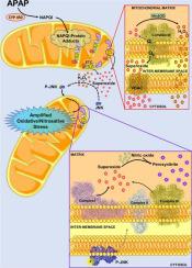
Acetaminophen (APAP) overdose is the most common cause of acute liver failure in the United States and formation of APAP-protein adducts, mitochondrial oxidant stress and activation of the mitogen activated protein (MAP) kinase c-jun N-terminal kinase (JNK) are critical for APAP-induced cell death. However, direct evidence linking these mechanistic features are lacking and were investigated by examining the early temporal course of these changes in mice after 300 mg/kg APAP. Protein adducts were detectable in the liver (0.05-0.1 nmol/mg protein) by 15 and 30 min after APAP, which increased (>500%) selectively in mitochondria by 60 min. Cytosolic JNK activation was only evident at 60 min, and was significantly attenuated by scavenging superoxide specifically in the cytosol by TEMPO treatment. Treatment of mouse hepatocytes with APAP revealed mitochondrial superoxide generation within 15 minutes, accompanied by hydrogen peroxide production without change in mitochondrial respiratory function. The oxidant stress preceded JNK activation and its mitochondrial translocation. Inhibitor studies identified the putative source of mitochondrial superoxide as complex III, which released superoxide towards the intermembrane space after APAP resulting in activation of JNK in the cytosol. Our studies provide direct evidence of mechanisms involved in mitochondrial superoxide generation after NAPQI-adduct formation and its activation of the MAP kinase cascade in the cytosol, which are critical features of APAP hepatotoxicity.
中文翻译:

线粒体蛋白加合物和超氧化物的产生是小鼠对乙酰氨基酚过量后早期激活细胞质内 c-Jun N-末端激酶的先决条件
对乙酰氨基酚 (APAP) 过量是美国急性肝衰竭和 APAP 蛋白加合物形成、线粒体氧化应激和丝裂原活化蛋白 (MAP) 激酶 c-jun N-末端激酶 (JNK) 激活的最常见原因对 APAP 诱导的细胞死亡至关重要。然而,缺乏将这些机械特征联系起来的直接证据,并通过检查 300 mg/kg APAP 后小鼠这些变化的早期时间过程进行了调查。APAP 后 15 分钟和 30 分钟可在肝脏中检测到蛋白质加合物(0.05-0.1 nmol/mg 蛋白质),60 分钟后在线粒体中选择性增加(>500%)。胞质 JNK 活化仅在 60 分钟时明显,并且通过 TEMPO 处理特异性清除胞质溶胶中的超氧化物显着减弱。用 APAP 处理小鼠肝细胞在 15 分钟内显示线粒体超氧化物的产生,伴随着过氧化氢的产生,而线粒体呼吸功能没有改变。氧化应激先于 JNK 活化及其线粒体易位。抑制剂研究确定了线粒体超氧化物的假定来源为复合物 III,其在 APAP 后向膜间隙释放超氧化物,导致胞质溶胶中 JNK 的激活。我们的研究直接证明了 NAPQI 加合物形成后线粒体超氧化物生成及其激活细胞质中 MAP 激酶级联的机制,这是 APAP 肝毒性的关键特征。伴随着过氧化氢的产生而不改变线粒体呼吸功能。氧化应激先于 JNK 活化及其线粒体易位。抑制剂研究确定了线粒体超氧化物的假定来源为复合物 III,其在 APAP 后向膜间隙释放超氧化物,导致胞质溶胶中 JNK 的激活。我们的研究直接证明了 NAPQI 加合物形成后线粒体超氧化物生成及其激活细胞质中 MAP 激酶级联的机制,这是 APAP 肝毒性的关键特征。伴随着过氧化氢的产生而不改变线粒体呼吸功能。氧化应激先于 JNK 活化及其线粒体易位。抑制剂研究确定线粒体超氧化物的假定来源为复合物 III,其在 APAP 后向膜间隙释放超氧化物,导致胞质溶胶中 JNK 的激活。我们的研究直接证明了 NAPQI 加合物形成后线粒体超氧化物生成及其激活细胞质中 MAP 激酶级联的机制,这是 APAP 肝毒性的关键特征。在 APAP 后向膜间隙释放超氧化物,导致胞质溶胶中 JNK 的激活。我们的研究直接证明了 NAPQI 加合物形成后线粒体超氧化物生成及其激活细胞质中 MAP 激酶级联的机制,这是 APAP 肝毒性的关键特征。在 APAP 后向膜间隙释放超氧化物,导致胞质溶胶中 JNK 的激活。我们的研究直接证明了 NAPQI 加合物形成后线粒体超氧化物生成及其激活细胞质中 MAP 激酶级联的机制,这是 APAP 肝毒性的关键特征。
更新日期:2021-03-01
中文翻译:

线粒体蛋白加合物和超氧化物的产生是小鼠对乙酰氨基酚过量后早期激活细胞质内 c-Jun N-末端激酶的先决条件
对乙酰氨基酚 (APAP) 过量是美国急性肝衰竭和 APAP 蛋白加合物形成、线粒体氧化应激和丝裂原活化蛋白 (MAP) 激酶 c-jun N-末端激酶 (JNK) 激活的最常见原因对 APAP 诱导的细胞死亡至关重要。然而,缺乏将这些机械特征联系起来的直接证据,并通过检查 300 mg/kg APAP 后小鼠这些变化的早期时间过程进行了调查。APAP 后 15 分钟和 30 分钟可在肝脏中检测到蛋白质加合物(0.05-0.1 nmol/mg 蛋白质),60 分钟后在线粒体中选择性增加(>500%)。胞质 JNK 活化仅在 60 分钟时明显,并且通过 TEMPO 处理特异性清除胞质溶胶中的超氧化物显着减弱。用 APAP 处理小鼠肝细胞在 15 分钟内显示线粒体超氧化物的产生,伴随着过氧化氢的产生,而线粒体呼吸功能没有改变。氧化应激先于 JNK 活化及其线粒体易位。抑制剂研究确定了线粒体超氧化物的假定来源为复合物 III,其在 APAP 后向膜间隙释放超氧化物,导致胞质溶胶中 JNK 的激活。我们的研究直接证明了 NAPQI 加合物形成后线粒体超氧化物生成及其激活细胞质中 MAP 激酶级联的机制,这是 APAP 肝毒性的关键特征。伴随着过氧化氢的产生而不改变线粒体呼吸功能。氧化应激先于 JNK 活化及其线粒体易位。抑制剂研究确定了线粒体超氧化物的假定来源为复合物 III,其在 APAP 后向膜间隙释放超氧化物,导致胞质溶胶中 JNK 的激活。我们的研究直接证明了 NAPQI 加合物形成后线粒体超氧化物生成及其激活细胞质中 MAP 激酶级联的机制,这是 APAP 肝毒性的关键特征。伴随着过氧化氢的产生而不改变线粒体呼吸功能。氧化应激先于 JNK 活化及其线粒体易位。抑制剂研究确定线粒体超氧化物的假定来源为复合物 III,其在 APAP 后向膜间隙释放超氧化物,导致胞质溶胶中 JNK 的激活。我们的研究直接证明了 NAPQI 加合物形成后线粒体超氧化物生成及其激活细胞质中 MAP 激酶级联的机制,这是 APAP 肝毒性的关键特征。在 APAP 后向膜间隙释放超氧化物,导致胞质溶胶中 JNK 的激活。我们的研究直接证明了 NAPQI 加合物形成后线粒体超氧化物生成及其激活细胞质中 MAP 激酶级联的机制,这是 APAP 肝毒性的关键特征。在 APAP 后向膜间隙释放超氧化物,导致胞质溶胶中 JNK 的激活。我们的研究直接证明了 NAPQI 加合物形成后线粒体超氧化物生成及其激活细胞质中 MAP 激酶级联的机制,这是 APAP 肝毒性的关键特征。


 京公网安备 11010802027423号