Experimental Eye Research ( IF 3.4 ) Pub Date : 2020-11-28 , DOI: 10.1016/j.exer.2020.108373 Neda Rashidi , Anup D. Pant , Samuel D. Salinas , Mickey Shah , Vineet S. Thomas , Ge Zhang , Syril Dorairaj , Rouzbeh Amini
|
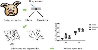
Nuclear shape alteration in ocular tissues, which can be used as a metric for overall cell deformation, may also lead to changes in gene expression and protein synthesis that could affect the biomechanics of the tissue extracellular matrix. The biomechanics of iris tissue is of particular interest in the study of primary angle-closure glaucoma. As the first step towards understanding the mutual role of the biomechanics and deformation of the iris on the activity of its constituent stromal cells, we conducted an ex-vivo study in freshly excised porcine eyes. Iris deformation was achieved by activating the constituent smooth muscles of the iris. Pupillary responses were initiated by inducing miosis and mydriasis, and the irides were placed in a fixative, bisected, and sliced into thin sections in a nasal and temporal horizontal orientation. The tissue sections were stained with DAPI for nucleus, and z-stacks were acquired using confocal microscopy. Images were analyzed to determine the nuclear aspect ratio (NAR) using both three-dimensional (3D) reconstructions of the nuclear surfaces as well as projections of the same 3D reconstruction into flat two-dimensional (2D) shapes. We observed that regardless of the calculation method (i.e., one that employed 3D surface reconstructions versus one that employed 2D projected images) the NAR increased in both the miosis group and the mydriasis group. Three-dimensional quantifications showed that NAR increased from 2.52 ± 0.96 in control group to 2.80 ± 0.81 and 2.74 ± 0.94 in the mydriasis and miosis groups, respectively. Notwithstanding the relative convenience in calculating the NAR using the 2D projected images, the 3D reconstructions were found to generate more physiologically realistic values and, thus, can be used in the development of future computational models to study primary angle-closure glaucoma. Since the iris undergoes large deformations in response to ambient light, this study suggests that the iris stromal cells are subjected to a biomechanically active micro-environment during their in-vivo physiological function.
中文翻译:
药理学诱发的瞳孔缩小和瞳孔散大时,虹膜基质细胞核变形为更细长的形状
眼组织中的核形状改变可用作整体细胞变形的度量,也可能导致基因表达和蛋白质合成发生变化,从而影响组织细胞外基质的生物力学。虹膜组织的生物力学在原发性闭角型青光眼的研究中尤为重要。作为了解虹膜生物力学和虹膜变形对其组成基质细胞活性的相互作用的第一步,我们进行了离体实验在刚摘下的猪眼中进行研究。虹膜变形是通过激活虹膜的组成平滑肌来实现的。瞳孔反应是通过诱导瞳孔缩小和瞳孔散大而引发的,并且将虹膜放入固定剂中,一分为二,并沿鼻和颞水平方向切成薄片。将组织切片用DAPI染色以检查细胞核,并使用共聚焦显微镜获得z堆栈。使用核表面的三维(3D)重建以及将相同3D重建投影到平坦的二维(2D)形状中的图像,分析图像以确定核的宽高比(NAR)。我们观察到,无论采用哪种计算方法(即 一种采用3D表面重建,另一种采用2D投影图像),在瞳孔缩小组和瞳孔散大组中,NAR均增加。三维量化显示,NAR从对照组的2.52±0.96增至散瞳和瞳孔缩小组的2.80±0.81和2.74±0.94。尽管使用2D投影图像计算NAR相对方便,但发现3D重建可产生更多的生理现实值,因此可用于将来的计算模型的开发中以研究原发性闭角型青光眼。由于虹膜在响应环境光时会发生较大的变形,因此这项研究表明虹膜基质细胞在其体内生理功能过程中会受到生物力学活性的微环境的影响。


 京公网安备 11010802027423号