当前位置:
X-MOL 学术
›
Ultrasonics
›
论文详情
Our official English website, www.x-mol.net, welcomes your feedback! (Note: you will need to create a separate account there.)
A fast technique for hyper-echoic region separation from brain ultrasound images using patch based thresholding and cubic B-spline based contour smoothing
Ultrasonics ( IF 4.2 ) Pub Date : 2021-03-01 , DOI: 10.1016/j.ultras.2020.106304 Haradhan Chel , P.K. Bora , K.K. Ramchiary
Ultrasonics ( IF 4.2 ) Pub Date : 2021-03-01 , DOI: 10.1016/j.ultras.2020.106304 Haradhan Chel , P.K. Bora , K.K. Ramchiary
|
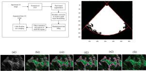
Ultrasound image guided brain surgery (UGBS) requires an automatic and fast image segmentation method. The level-set and active contour based algorithms have been found to be useful for obtaining topology-independent boundaries between different image regions. But slow convergence limits their use in online US image segmentation. The performance of these algorithms deteriorates on US images because of the intensity inhomogeneity. This paper proposes an effective region-driven method for the segmentation of hyper-echoic (HE) regions suppressing the hypo-echoic and anechoic regions in brain US images. An automatic threshold estimation scheme is developed with a modified Niblack's approach. The separation of the hyper-echoic and non-hyper-echoic (NHE) regions is performed by successively applying patch based intensity thresholding and boundary smoothing. First, a patch based segmentation is performed, which separates roughly the two regions. The patch based approach in this process reduces the effect of intensity heterogeneity within an HE region. An iterative boundary correction step with reducing patch size improves further the regional topology and refines the boundary regions. For avoiding the slope and curvature discontinuities and obtaining distinct boundaries between HE and NHE regions, a cubic B-spline model of curve smoothing is applied. The proposed method is 50-100 times faster than the other level-set based image segmentation algorithms. The segmentation performance and the convergence speed of the proposed method are compared with four other competing level-set based algorithms. The computational results show that the proposed segmentation approach outperforms other level-set based techniques both subjectively and objectively.
中文翻译:

一种使用基于补丁的阈值和基于三次 B 样条的轮廓平滑从脑超声图像中分离高回声区域的快速技术
超声图像引导脑外科手术 (UGBS) 需要一种自动且快速的图像分割方法。已经发现基于水平集和活动轮廓的算法可用于获得不同图像区域之间的拓扑独立边界。但是缓慢的收敛限制了它们在在线美国图像分割中的使用。由于强度不均匀,这些算法在美国图像上的性能下降。本文提出了一种有效的区域驱动方法,用于分割高回声 (HE) 区域,以抑制大脑超声图像中的低回声和无回声区域。使用改进的 Niblack 方法开发了自动阈值估计方案。高回声和非高回声 (NHE) 区域的分离是通过连续应用基于补丁的强度阈值和边界平滑来执行的。首先,执行基于补丁的分割,大致将两个区域分开。该过程中基于补丁的方法减少了 HE 区域内强度异质性的影响。减少补丁大小的迭代边界校正步骤进一步改进了区域拓扑并细化了边界区域。为了避免斜率和曲率不连续性并获得 HE 和 NHE 区域之间的明显边界,应用了曲线平滑的三次 B 样条模型。所提出的方法比其他基于水平集的图像分割算法快 50-100 倍。将所提出方法的分割性能和收敛速度与其他四种基于水平集的竞争算法进行了比较。计算结果表明,所提出的分割方法在主观和客观上都优于其他基于水平集的技术。
更新日期:2021-03-01
中文翻译:
一种使用基于补丁的阈值和基于三次 B 样条的轮廓平滑从脑超声图像中分离高回声区域的快速技术
超声图像引导脑外科手术 (UGBS) 需要一种自动且快速的图像分割方法。已经发现基于水平集和活动轮廓的算法可用于获得不同图像区域之间的拓扑独立边界。但是缓慢的收敛限制了它们在在线美国图像分割中的使用。由于强度不均匀,这些算法在美国图像上的性能下降。本文提出了一种有效的区域驱动方法,用于分割高回声 (HE) 区域,以抑制大脑超声图像中的低回声和无回声区域。使用改进的 Niblack 方法开发了自动阈值估计方案。高回声和非高回声 (NHE) 区域的分离是通过连续应用基于补丁的强度阈值和边界平滑来执行的。首先,执行基于补丁的分割,大致将两个区域分开。该过程中基于补丁的方法减少了 HE 区域内强度异质性的影响。减少补丁大小的迭代边界校正步骤进一步改进了区域拓扑并细化了边界区域。为了避免斜率和曲率不连续性并获得 HE 和 NHE 区域之间的明显边界,应用了曲线平滑的三次 B 样条模型。所提出的方法比其他基于水平集的图像分割算法快 50-100 倍。将所提出方法的分割性能和收敛速度与其他四种基于水平集的竞争算法进行了比较。计算结果表明,所提出的分割方法在主观和客观上都优于其他基于水平集的技术。


 京公网安备 11010802027423号