Acta Biomaterialia ( IF 9.7 ) Pub Date : 2020-11-21 , DOI: 10.1016/j.actbio.2020.11.028 Marley J Dewey 1 , Andrey V Nosatov 1 , Kiran Subedi 2 , Ramille Shah 3 , Adam Jakus 3 , Brendan A C Harley 4
|
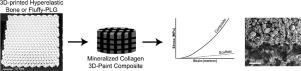
Regenerative repair of craniomaxillofacial bone injuries is challenging due to both the large size and irregular shape of many defects. Mineralized collagen scaffolds have previously been shown to be a promising biomaterial implant to accelerate craniofacial bone regeneration in vivo. Here we describe inclusion of a 3D-printed polymer or ceramic-based mesh into a mineralized collagen scaffold to improve mechanical and biological activity. Mineralized collagen scaffolds were reinforced with 3D-printed Fluffy-PLG (ultraporous polylactide-co-glycolide co-polymer) or Hyperelastic Bone (90wt% calcium phosphate in PLG) meshes. We show degradation byproducts and acidic release from the printed structures have limited negative impact on the viability of mesenchymal stem cells. Further, inclusion of a mesh formed from Hyperelastic Bone generates a reinforced composite with significantly improved mechanical performance (elastic modulus, push-out strength). Composites formed from the mineralized collagen scaffold and either Hyperelastic Bone or Fluffy-PLG reinforcement both supported human bone-marrow derived mesenchymal stem cell osteogenesis and new bone formation. This was observed by increased mineral formation in Fluffy-PLG composites and increased cell viability and upregulation of RUNX2, Osterix, and COL1A2 genes in both composites. Strikingly, composites reinforced with Hyperelastic Bone mesh elicited significantly increased secretion of osteoprotegerin, a soluble glycoprotein and endogenous inhibitor of osteoclast activity. These results suggest that architectured meshes can be integrated into collagen scaffolds to boost mechanical performance and actively instruct cell processes that aid osteogenicity; specifically, secretion of a factor crucial to inhibiting osteoclast-mediated bone resorption. Future work will focus on further adapting the polymer mesh architecture to confer improved shape-fitting capacity as well as to investigate the role of polymer reinforcement on MSC-osteoclast interactions as a means to increase regenerative potential.
中文翻译:
包含 3D 打印的超弹性骨网可改善矿化胶原支架的机械和成骨性能
由于许多缺陷的尺寸较大且形状不规则,颅颌面骨损伤的再生修复具有挑战性。矿化胶原支架先前已被证明是一种有前途的生物材料植入物,可在体内加速颅面骨再生. 在这里,我们描述了在矿化胶原支架中加入 3D 打印聚合物或陶瓷网,以提高机械和生物活性。矿化胶原蛋白支架用 3D 打印的 Fluffy-PLG(超多孔聚乳酸-乙交酯共聚物)或超弹性骨(PLG 中 90wt% 的磷酸钙)网格进行加固。我们表明,印刷结构的降解副产物和酸性释放对间充质干细胞活力的负面影响有限。此外,包含由超弹性骨形成的网格会产生具有显着改善的机械性能(弹性模量、推出强度)的增强复合材料。由矿化胶原支架和超弹性骨或 Fluffy-PLG 增强材料形成的复合材料都支持人类骨髓来源的间充质干细胞成骨和新骨形成。这是通过在 Fluffy-PLG 复合材料中增加矿物质形成和增加细胞活力和上调两种复合材料中的RUNX2、Osterix和COL1A2基因。引人注目的是,用超弹性骨网增强的复合材料显着增加了骨保护素的分泌,骨保护素是一种可溶性糖蛋白和破骨细胞活性的内源性抑制剂。这些结果表明,结构化网格可以整合到胶原蛋白支架中,以提高机械性能并积极指导有助于成骨的细胞过程;具体来说,分泌一种对抑制破骨细胞介导的骨吸收至关重要的因子。未来的工作将集中在进一步调整聚合物网格结构以提高形状拟合能力,并研究聚合物增强在 MSC-破骨细胞相互作用中的作用,作为增加再生潜力的一种手段。


 京公网安备 11010802027423号