当前位置:
X-MOL 学术
›
Mol. Pharmaceutics
›
论文详情
Our official English website, www.x-mol.net, welcomes your feedback! (Note: you will need to create a separate account there.)
[18F]-FMISO PET/MRI Imaging Shows Ischemic Tissue around Hematoma in Intracerebral Hemorrhage
Molecular Pharmaceutics ( IF 4.9 ) Pub Date : 2020-11-13 , DOI: 10.1021/acs.molpharmaceut.0c00932 Noemí Gómez-Lado 1, 2 , Esteban López-Arias 3 , Ramón Iglesias-Rey 3 , Lucía Díaz-Platas 4 , Santiago Medín-Aguerre 4 , Anxo Fernández-Ferreiro 5 , Adrián Posado-Fernández 3 , Lara García-Varela 1 , Manuel Rodríguez-Pérez 3 , Francisco Campos 3 , Pablo Del Pino 6 , Álvaro Ruibal 1, 2 , Juan Pardo-Montero 7 , José Castillo 3 , Pablo Aguiar 1, 2 , Tomás Sobrino 3
Molecular Pharmaceutics ( IF 4.9 ) Pub Date : 2020-11-13 , DOI: 10.1021/acs.molpharmaceut.0c00932 Noemí Gómez-Lado 1, 2 , Esteban López-Arias 3 , Ramón Iglesias-Rey 3 , Lucía Díaz-Platas 4 , Santiago Medín-Aguerre 4 , Anxo Fernández-Ferreiro 5 , Adrián Posado-Fernández 3 , Lara García-Varela 1 , Manuel Rodríguez-Pérez 3 , Francisco Campos 3 , Pablo Del Pino 6 , Álvaro Ruibal 1, 2 , Juan Pardo-Montero 7 , José Castillo 3 , Pablo Aguiar 1, 2 , Tomás Sobrino 3
Affiliation

|
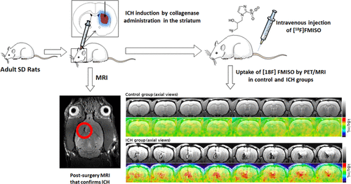
Intracerebral hemorrhage (ICH), being the most severe cerebrovascular disease, accounts for 10–15% of all strokes. Hematoma expansion is one of the most important factors associated with poor outcome in intracerebral hemorrhage (ICH). Several studies have suggested that an “ischemic penumbra” might arise when the hematoma has a large expansion, but clinical studies are inconclusive. We performed a preclinical study to demonstrate the presence of hypoxic–ischemic tissue around the hematoma by means of longitudinal [18F]-fluoromisonidazole ([18F]-FMISO) PET/MRI studies over time in an experimental ICH model. Our results showed that all [18F]-FMISO PET/MRI images exhibited hypoxic–ischemic tissue around the hematoma area. A significant increase of [18F]-FMISO uptake was found at 18–24 h post-ICH when the maximum of hematoma volume is achieved and this increase disappeared before 42 h. These results demonstrate the presence of hypoxic tissue around the hematoma and open the possibility of new therapies aimed to reduce ischemic damage associated with ICH.
中文翻译:

[18F]-FMISO PET/MRI 成像显示脑出血血肿周围的缺血组织
脑出血(ICH)是最严重的脑血管疾病,占所有卒中的 10-15%。血肿扩大是与脑出血 (ICH) 预后不良相关的最重要因素之一。多项研究表明,当血肿大范围扩张时,可能会出现“缺血半影区”,但临床研究尚无定论。我们进行了一项临床前研究,通过纵向 [ 18 F]-氟咪唑([ 18 F]-FMISO) PET/MRI 研究在实验性 ICH 模型中随时间推移证明血肿周围存在缺氧缺血组织。我们的结果表明,所有[ 18 F]-FMISO PET/MRI 图像都显示血肿区域周围的缺氧缺血组织。显着增加 [18 F]-FMISO 在 ICH 后 18-24 小时被发现,此时血肿体积达到最大,这种增加在 42 小时前消失。这些结果表明血肿周围存在缺氧组织,并开启了旨在减少与 ICH 相关的缺血性损伤的新疗法的可能性。
更新日期:2020-12-07
中文翻译:

[18F]-FMISO PET/MRI 成像显示脑出血血肿周围的缺血组织
脑出血(ICH)是最严重的脑血管疾病,占所有卒中的 10-15%。血肿扩大是与脑出血 (ICH) 预后不良相关的最重要因素之一。多项研究表明,当血肿大范围扩张时,可能会出现“缺血半影区”,但临床研究尚无定论。我们进行了一项临床前研究,通过纵向 [ 18 F]-氟咪唑([ 18 F]-FMISO) PET/MRI 研究在实验性 ICH 模型中随时间推移证明血肿周围存在缺氧缺血组织。我们的结果表明,所有[ 18 F]-FMISO PET/MRI 图像都显示血肿区域周围的缺氧缺血组织。显着增加 [18 F]-FMISO 在 ICH 后 18-24 小时被发现,此时血肿体积达到最大,这种增加在 42 小时前消失。这些结果表明血肿周围存在缺氧组织,并开启了旨在减少与 ICH 相关的缺血性损伤的新疗法的可能性。


 京公网安备 11010802027423号