当前位置:
X-MOL 学术
›
Ann. N. Y. Acad. Sci.
›
论文详情
Our official English website, www.x-mol.net, welcomes your feedback! (Note: you will need to create a separate account there.)
Advanced endoscopic imaging for detecting and guiding therapy of early neoplasias of the esophagus
Annals of the New York Academy of Sciences ( IF 5.2 ) Pub Date : 2020-11-12 , DOI: 10.1111/nyas.14523 Hiroshi Mashimo 1, 2 , Stuart R Gordon 3 , Satish K Singh 1, 4
Annals of the New York Academy of Sciences ( IF 5.2 ) Pub Date : 2020-11-12 , DOI: 10.1111/nyas.14523 Hiroshi Mashimo 1, 2 , Stuart R Gordon 3 , Satish K Singh 1, 4
Affiliation
|
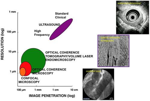
Esophageal cancers, largely adenocarcinoma in Western countries and squamous cell cancer in Asia, present a significant burden of disease and remain one of the most lethal of cancers. Key to improving survival is the development and adoption of new imaging modalities to identify early neoplastic lesions, which may be small, multifocal, subsurface, and difficult to detect by standard endoscopy. Such advanced imaging is particularly relevant with the emergence of ablative techniques that often require multiple endoscopic sessions and may be complicated by bleeding, pain, strictures, and recurrences. Assessing the specific location, depth of involvement, and features correlated with neoplastic progression or incomplete treatment may optimize treatments. While not comprehensive of all endoscopic imaging modalities, we review here some of the recent advances in endoscopic luminal imaging, particularly with surface contrast enhancement using virtual chromoendoscopy, highly magnified subsurface imaging with confocal endomicroscopy, optical coherence tomography, elastic scattering spectroscopy, angle‐resolved low‐coherence interferometry, and light scattering spectroscopy. While there is no single ideal imaging modality, various multimodal instruments are also being investigated. The future of combining computer‐aided assessments, molecular markers, and improved imaging technologies to help localize and ablate early neoplastic lesions shed hope for improved disease outcome.
中文翻译:

用于检测和指导食管早期肿瘤的先进内窥镜成像
食道癌,主要是西方国家的腺癌和亚洲的鳞状细胞癌,造成了重大的疾病负担,并且仍然是最致命的癌症之一。提高生存率的关键是开发和采用新的成像方式来识别早期肿瘤病变,这些病变可能很小、多灶性、次表面,并且难以通过标准内窥镜检查发现。这种先进的成像与消融技术的出现特别相关,这些技术通常需要多次内窥镜检查,并且可能因出血、疼痛、狭窄和复发而变得复杂。评估与肿瘤进展或不完全治疗相关的特定位置、受累深度和特征可以优化治疗。虽然并非所有内窥镜成像方式都全面,我们在这里回顾了内窥镜管腔成像的一些最新进展,特别是使用虚拟色素内窥镜的表面对比度增强、使用共焦内窥镜的高放大次表面成像、光学相干断层扫描、弹性散射光谱、角分辨低相干干涉测量和光散射光谱. 虽然没有单一的理想成像模式,但也正在研究各种多模式仪器。结合计算机辅助评估、分子标记和改进的成像技术来帮助定位和消融早期肿瘤病变的未来为改善疾病结果带来了希望。光学相干断层扫描、弹性散射光谱、角分辨低相干干涉测量和光散射光谱。虽然没有单一的理想成像模式,但也正在研究各种多模式仪器。结合计算机辅助评估、分子标记和改进的成像技术来帮助定位和消融早期肿瘤病变的未来为改善疾病结果带来了希望。光学相干断层扫描、弹性散射光谱、角分辨低相干干涉测量和光散射光谱。虽然没有单一的理想成像模式,但也正在研究各种多模式仪器。结合计算机辅助评估、分子标记和改进的成像技术来帮助定位和消融早期肿瘤病变的未来为改善疾病结果带来了希望。
更新日期:2020-11-12
中文翻译:
用于检测和指导食管早期肿瘤的先进内窥镜成像
食道癌,主要是西方国家的腺癌和亚洲的鳞状细胞癌,造成了重大的疾病负担,并且仍然是最致命的癌症之一。提高生存率的关键是开发和采用新的成像方式来识别早期肿瘤病变,这些病变可能很小、多灶性、次表面,并且难以通过标准内窥镜检查发现。这种先进的成像与消融技术的出现特别相关,这些技术通常需要多次内窥镜检查,并且可能因出血、疼痛、狭窄和复发而变得复杂。评估与肿瘤进展或不完全治疗相关的特定位置、受累深度和特征可以优化治疗。虽然并非所有内窥镜成像方式都全面,我们在这里回顾了内窥镜管腔成像的一些最新进展,特别是使用虚拟色素内窥镜的表面对比度增强、使用共焦内窥镜的高放大次表面成像、光学相干断层扫描、弹性散射光谱、角分辨低相干干涉测量和光散射光谱. 虽然没有单一的理想成像模式,但也正在研究各种多模式仪器。结合计算机辅助评估、分子标记和改进的成像技术来帮助定位和消融早期肿瘤病变的未来为改善疾病结果带来了希望。光学相干断层扫描、弹性散射光谱、角分辨低相干干涉测量和光散射光谱。虽然没有单一的理想成像模式,但也正在研究各种多模式仪器。结合计算机辅助评估、分子标记和改进的成像技术来帮助定位和消融早期肿瘤病变的未来为改善疾病结果带来了希望。光学相干断层扫描、弹性散射光谱、角分辨低相干干涉测量和光散射光谱。虽然没有单一的理想成像模式,但也正在研究各种多模式仪器。结合计算机辅助评估、分子标记和改进的成像技术来帮助定位和消融早期肿瘤病变的未来为改善疾病结果带来了希望。


 京公网安备 11010802027423号