当前位置:
X-MOL 学术
›
ACS Photonics
›
论文详情
Our official English website, www.x-mol.net, welcomes your feedback! (Note: you will need to create a separate account there.)
Guided-Mode-Resonant Dielectric Metasurfaces for Colorimetric Imaging of Material Anisotropy in Fibrous Biological Tissue
ACS Photonics ( IF 7 ) Pub Date : 2020-11-05 , DOI: 10.1021/acsphotonics.0c01303 Lisa V. Poulikakos 1, 2 , Mark Lawrence 1 , David R. Barton 1 , Stefanie S. Jeffrey 3 , Jennifer A. Dionne 1
ACS Photonics ( IF 7 ) Pub Date : 2020-11-05 , DOI: 10.1021/acsphotonics.0c01303 Lisa V. Poulikakos 1, 2 , Mark Lawrence 1 , David R. Barton 1 , Stefanie S. Jeffrey 3 , Jennifer A. Dionne 1
Affiliation
|
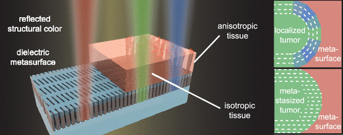
The structural arrangement of fibrous tissue is linked to the onset and progression of Alzheimer’s disease, heart disease, fibrosis, and cancer, yet its visualization remains challenging with conventional optical microscopy. Here, we design a guided-mode-resonant dielectric metasurface to detect the presence and orientation of fibrous tissue, modeled as a linearly birefringent anisotropic medium, by colorimetric readout. The metasurface consists of nanoscale Si3N4 (215 nm) and SiO2 (75 nm) layers on a SiO2 substrate, acting as a broadband antireflection coating, patterned with subwavelength-periodic rhombohedral perturbations, which result in guided-mode resonances with sub-10 nm bandwidth. Using full-field simulations, we show how transition from air to tissue in the dielectric environment at the metasurface interface results in a red-to-green change in reflected structural color, while the birefringence and orientation of an anisotropic medium manifests as a green-to-blue change. Importantly, the birefringence-based tuning of the guided mode resonances is spectrally separated from refractive-index-based displacements, allowing quantitative discrimination between both the index and structural arrangement of anisotropic media. We numerically simulate the application of our metasurface to cancer tissue diagnostics, predicting how changes in reflected structural color at the tumor margin can distinguish localized, early stage from metastasized, late-stage cancers. The quantitative, colorimetric mapping of tissue orientation angle marks an improved performance in comparison to polarized light microscopy, where multiple orientation angles yield an identical response. Our guided-mode-resonant metasurface provides a foundation for all-optical, label-free, and quantitative colorimetric visualization of fibrous biological media on a single, clinically compatible chip, promising improvements in staging and treatment decisions.
中文翻译:

导模共振介电超表面用于纤维生物组织中材料各向异性的比色成像。
纤维组织的结构排列与阿尔茨海默氏病,心脏病,纤维化和癌症的发生和发展有关,但是其可视化仍然是传统光学显微镜所面临的挑战。在这里,我们设计了一种导模共振介电超表面,以通过比色法读出来检测纤维组织的存在和取向,该组织被建模为线性双折射各向异性介质。超颖表面包括纳米尺度的Si 3 Ñ 4(215纳米)和SiO 2(75 nm)的层上形成SiO 2基板,用作宽带抗反射涂层,具有亚波长周期的菱面体微扰,可在低于10 nm的带宽下产生导模共振。使用全场模拟,我们显示出在超表面界面处介电环境中从空气到组织的过渡如何导致反射的结构颜色由红变绿,而各向异性介质的双折射和取向则表现为绿色。变成蓝色。重要的是,引导模式共振的基于双折射的调谐与基于折射率的位移在光谱上是分开的,从而可以定量区分各向异性介质的折射率和结构。我们通过数值模拟我们的超表面在癌症组织诊断中的应用,预测肿瘤边缘反射结构颜色的变化如何区分局部早期和转移性晚期癌症。与偏光显微镜相比,组织取向角的定量,比色映射标志着性能的提高,在偏振光显微镜中,多个取向角产生相同的响应。我们的导模共振超表面为在临床上兼容的单个芯片上对纤维生物介质进行全光学,无标签和定量比色可视化提供了基础,有望改善分期和治疗决策。组织取向角的比色映射与偏振光显微镜相比具有更好的性能,在偏振光显微镜中,多个取向角产生相同的响应。我们的导模共振超表面为在临床上兼容的单个芯片上对纤维生物介质进行全光学,无标签和定量比色可视化提供了基础,有望改善分期和治疗决策。组织取向角的比色映射与偏振光显微镜相比具有更好的性能,在偏振光显微镜中,多个取向角产生相同的响应。我们的引导模式共振超表面为在临床上兼容的单个芯片上对纤维生物介质进行全光学,无标记和定量比色可视化提供了基础,有望改善分期和治疗决策。
更新日期:2020-11-18
中文翻译:
导模共振介电超表面用于纤维生物组织中材料各向异性的比色成像。
纤维组织的结构排列与阿尔茨海默氏病,心脏病,纤维化和癌症的发生和发展有关,但是其可视化仍然是传统光学显微镜所面临的挑战。在这里,我们设计了一种导模共振介电超表面,以通过比色法读出来检测纤维组织的存在和取向,该组织被建模为线性双折射各向异性介质。超颖表面包括纳米尺度的Si 3 Ñ 4(215纳米)和SiO 2(75 nm)的层上形成SiO 2基板,用作宽带抗反射涂层,具有亚波长周期的菱面体微扰,可在低于10 nm的带宽下产生导模共振。使用全场模拟,我们显示出在超表面界面处介电环境中从空气到组织的过渡如何导致反射的结构颜色由红变绿,而各向异性介质的双折射和取向则表现为绿色。变成蓝色。重要的是,引导模式共振的基于双折射的调谐与基于折射率的位移在光谱上是分开的,从而可以定量区分各向异性介质的折射率和结构。我们通过数值模拟我们的超表面在癌症组织诊断中的应用,预测肿瘤边缘反射结构颜色的变化如何区分局部早期和转移性晚期癌症。与偏光显微镜相比,组织取向角的定量,比色映射标志着性能的提高,在偏振光显微镜中,多个取向角产生相同的响应。我们的导模共振超表面为在临床上兼容的单个芯片上对纤维生物介质进行全光学,无标签和定量比色可视化提供了基础,有望改善分期和治疗决策。组织取向角的比色映射与偏振光显微镜相比具有更好的性能,在偏振光显微镜中,多个取向角产生相同的响应。我们的导模共振超表面为在临床上兼容的单个芯片上对纤维生物介质进行全光学,无标签和定量比色可视化提供了基础,有望改善分期和治疗决策。组织取向角的比色映射与偏振光显微镜相比具有更好的性能,在偏振光显微镜中,多个取向角产生相同的响应。我们的引导模式共振超表面为在临床上兼容的单个芯片上对纤维生物介质进行全光学,无标记和定量比色可视化提供了基础,有望改善分期和治疗决策。


 京公网安备 11010802027423号