Biomaterials Advances ( IF 7.9 ) Pub Date : 2020-11-06 , DOI: 10.1016/j.msec.2020.111707 Omar A. Hamid , Hoda M. Eltaher , Virginie Sottile , Jing Yang
|
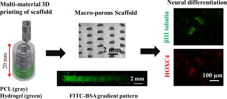
Development of a biomimetic tubular scaffold capable of recreating developmental neurogenesis using pluripotent stem cells offers a novel strategy for the repair of spinal cord tissues. Recent advances in 3D printing technology have facilitated biofabrication of complex biomimetic environments by precisely controlling the 3D arrangement of various acellular and cellular components (biomaterials, cells and growth factors). Here, we present a 3D printing method to fabricate a complex, patterned and embryoid body (EB)-laden tubular scaffold composed of polycaprolactone (PCL) and hydrogel (alginate or gelatine methacrylate (GelMA)). Our results revealed 3D printing of a strong, macro-porous PCL/hydrogel tubular scaffold with a high capacity to control the porosity of the PCL scaffold, wherein the maximum porosity in the PCL wall was 15%. The method was equally employed to create spatiotemporal protein concentration within the scaffold, demonstrating its ability to generate linear and opposite gradients of model molecules (fluorescein isothiocyanate-conjugated bovine serum albumin (FITC-BSA) and rhodamine). 3D bioprinting of EBs-laden GelMA was introduced as a novel 3D printing strategy to incorporate EBs in a hydrogel matrix. Cell viability and proliferation were measured post-printing. Following the bioprinting of EBs-laden 5% GelMA hydrogel, neural differentiation of EBs was induced using 1 μM retinoic acid (RA). The differentiated EBs contained βIII-tubulin positive neurons displaying axonal extensions and cells migration. Finally, 3D bioprinting of EBs-laden PCL/GelMA tubular scaffold successfully supported EBs neural differentiation and patterning in response to co-printing with 1 μM RA. 3D printing of a complex heterogeneous tubular scaffold that can encapsulate EBs, spatially controlled protein concentration and promote neuronal patterning will help in developing more biomimetic scaffolds capable of replicating the neural patterning which occurs during neural tube development.
中文翻译:
充满干细胞的多材料管状复合材料的3D生物打印:脊髓修复的方法
能够使用多能干细胞重建仿生管状支架的仿生管状支架的开发,为修复脊髓组织提供了一种新的策略。3D打印技术的最新进展通过精确控制各种脱细胞和细胞成分(生物材料,细胞和生长因子)的3D排列,促进了复杂仿生环境的生物制造。在这里,我们提出了一种3D打印方法来制造载有聚己内酯(PCL)和水凝胶(藻酸盐或甲基丙烯酸明胶(GelMA))的复杂,有图案且具胚状体(EB)的管状支架。我们的结果表明,坚固,大孔的PCL /水凝胶管状支架的3D打印具有很高的控制PCL支架孔隙率的能力,其中PCL壁中的最大孔隙率为15%。该方法同样用于在支架中产生时空蛋白浓度,证明其能够产生线性和相反梯度的模型分子(异硫氰酸荧光素缀合的牛血清白蛋白(FITC-BSA)和若丹明)。载有EB的GelMA的3D生物打印是一种新颖的3D打印策略,用于将EB掺入水凝胶基质中。在印刷后测量细胞活力和增殖。在载有EB的5%GelMA水凝胶进行生物打印后,使用1μM视黄酸(RA)诱导EB的神经分化。分化的EBs包含βIII-微管蛋白阳性神经元,表现出轴突延伸和细胞迁移。最后,载有EB的PCL / GelMA管状支架的3D生物打印成功支持了EB的神经分化和模式响应,与1μMRA共同印刷。复杂的异质管状支架的3D打印可以封装EB,在空间上控制蛋白质的浓度并促进神经元模式的形成,将有助于开发更多的仿生支架,从而能够复制在神经管发育过程中发生的神经模式。


 京公网安备 11010802027423号