当前位置:
X-MOL 学术
›
Mol. Pharmaceutics
›
论文详情
Our official English website, www.x-mol.net, welcomes your feedback! (Note: you will need to create a separate account there.)
Disparities of Single-Particle Growth Rates in Buried Versus Exposed Ritonavir Crystals within Amorphous Solid Dispersions
Molecular Pharmaceutics ( IF 4.9 ) Pub Date : 2020-11-05 , DOI: 10.1021/acs.molpharmaceut.0c00744 Scott R Griffin 1 , Nita Takanti 1 , Sreya Sarkar 1 , Zhengtian Song 1 , Andrew D Vogt 2 , Gerald D Danzer 2 , Garth J Simpson 1
Molecular Pharmaceutics ( IF 4.9 ) Pub Date : 2020-11-05 , DOI: 10.1021/acs.molpharmaceut.0c00744 Scott R Griffin 1 , Nita Takanti 1 , Sreya Sarkar 1 , Zhengtian Song 1 , Andrew D Vogt 2 , Gerald D Danzer 2 , Garth J Simpson 1
Affiliation
|
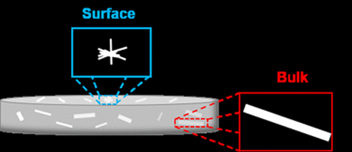
Seeded growth rates of ritonavir in copovidone at 75% relative humidity (RH) and 50 °C were evaluated by single-particle tracking second harmonic generation (SHG) microscopy and found to be ∼3-fold slower for crystallites at the surface compared to the bulk. The shelf lives of final dosage forms containing amorphous solid dispersions (ASDs) are often dictated by the rates of active pharmaceutical ingredient crystallization. Upon exposure to elevated RH, the higher anticipated water content near the surfaces of ASDs has the potential to substantially impact nucleation and growth kinetics relative to the bulk. However, quantitative assessment of these differences in growth rates is complicated by challenges associated with discrimination of the two contributions (supersaturation and molecular mobility) in ensemble-averaged measurements. In the present study, “sandwich” materials were prepared, in which sparse populations of ritonavir single-crystalline seeds were pressed between two similar ASD films to assess bulk crystallization rates. These sandwich materials were compared and contrasted with analogously prepared “open-faced” samples, without the capping film, to assess the surface crystallization rates. Single-particle analysis by SHG microscopy time-series during in situ crystallization produced average growth rates of 3.8 μm/h for bulk columnar crystals with a particle-to-particle standard deviation of 0.9 μm/h. In addition, columnar crystal growth rates for surface particles were measured to be 1.3 μm/h and radiating crystal growth rates for surface particles were measured to be 1.0 μm/h, both with a particle-to-particle deviation of 0.4 μm/h. The observed appearance of radiating crystals upon surface seeding is attributed to reduced ritonavir solubility upon water adsorption at the interface, leading to higher defect densities in crystal growth. Despite substantial differences in crystal habit, correction of the surface growth rates by a factor of 4 from geometric effects resulted in relatively minor but statistically significant differences in the growth kinetics for the two local environments. These results are consistent, with viscosity being a relatively weak function of water absorption coupled with primarily diffusion-limited growth kinetics.
中文翻译:

无定形固体分散体中埋藏与暴露的利托那韦晶体中单粒子生长速率的差异
在 75% 相对湿度 (RH) 和 50 °C 条件下,利托那韦在共聚维酮中的种子生长速率通过单粒子跟踪二次谐波发生 (SHG) 显微镜进行评估,发现与表面微晶相比,表面微晶慢约 3 倍。大部分。含有无定形固体分散体 (ASD) 的最终剂型的保质期通常取决于活性药物成分的结晶速率。在暴露于升高的 RH 时,ASD 表面附近较高的预期含水量有可能显着影响相对于整体的成核和生长动力学。然而,由于在整体平均测量中区分两种贡献(过饱和和分子迁移率)相关的挑战,对这些增长率差异的定量评估变得复杂。在本研究中,制备了“夹心”材料,其中将稀疏的利托那韦单晶种子压在两个相似的 ASD 薄膜之间,以评估整体结晶速率。将这些夹心材料与类似制备的“开放面”样品进行比较和对比,没有覆盖膜,以评估表面结晶速率。SHG 显微镜下的单粒子分析时间序列就地结晶使块状柱状晶体的平均生长速率为 3.8 μm/h,颗粒间标准偏差为 0.9 μm/h。此外,表面颗粒的柱状晶体生长速率测量为 1.3 μm/h,表面颗粒的辐射晶体生长速率测量为 1.0 μm/h,两者的颗粒间偏差均为 0.4 μm/h。在表面接种时观察到的辐射晶体的出现归因于在界面处吸附水时利托那韦的溶解度降低,导致晶体生长中更高的缺陷密度。尽管晶体习性存在显着差异,但根据几何效应将表面生长速率校正为 4 倍,导致两种局部环境的生长动力学差异相对较小但具有统计学意义。
更新日期:2020-12-07
中文翻译:
无定形固体分散体中埋藏与暴露的利托那韦晶体中单粒子生长速率的差异
在 75% 相对湿度 (RH) 和 50 °C 条件下,利托那韦在共聚维酮中的种子生长速率通过单粒子跟踪二次谐波发生 (SHG) 显微镜进行评估,发现与表面微晶相比,表面微晶慢约 3 倍。大部分。含有无定形固体分散体 (ASD) 的最终剂型的保质期通常取决于活性药物成分的结晶速率。在暴露于升高的 RH 时,ASD 表面附近较高的预期含水量有可能显着影响相对于整体的成核和生长动力学。然而,由于在整体平均测量中区分两种贡献(过饱和和分子迁移率)相关的挑战,对这些增长率差异的定量评估变得复杂。在本研究中,制备了“夹心”材料,其中将稀疏的利托那韦单晶种子压在两个相似的 ASD 薄膜之间,以评估整体结晶速率。将这些夹心材料与类似制备的“开放面”样品进行比较和对比,没有覆盖膜,以评估表面结晶速率。SHG 显微镜下的单粒子分析时间序列就地结晶使块状柱状晶体的平均生长速率为 3.8 μm/h,颗粒间标准偏差为 0.9 μm/h。此外,表面颗粒的柱状晶体生长速率测量为 1.3 μm/h,表面颗粒的辐射晶体生长速率测量为 1.0 μm/h,两者的颗粒间偏差均为 0.4 μm/h。在表面接种时观察到的辐射晶体的出现归因于在界面处吸附水时利托那韦的溶解度降低,导致晶体生长中更高的缺陷密度。尽管晶体习性存在显着差异,但根据几何效应将表面生长速率校正为 4 倍,导致两种局部环境的生长动力学差异相对较小但具有统计学意义。


 京公网安备 11010802027423号