当前位置:
X-MOL 学术
›
Brain Pathol.
›
论文详情
Our official English website, www.x-mol.net, welcomes your feedback! (Note: you will need to create a separate account there.)
Intracranial mesenchymal tumor with FET-CREB fusion—A unifying diagnosis for the spectrum of intracranial myxoid mesenchymal tumors and angiomatoid fibrous histiocytoma-like neoplasms
Brain Pathology ( IF 6.4 ) Pub Date : 2020-11-03 , DOI: 10.1111/bpa.12918 Emily A Sloan 1 , Jason Chiang 2 , Javier E Villanueva-Meyer 3 , Sanda Alexandrescu 4 , Jennifer M Eschbacher 5 , Wesley Wang 6 , Manuela Mafra 7 , Nasir Ud Din 8 , Emily Carr-Boyd 9 , Michael Watson 9 , Michael Punsoni 10 , Angelica Oviedo 11 , Ahmed Gilani 12 , Bette K Kleinschmidt-DeMasters 12 , Dylan J Coss 13 , M Beatriz Lopes 13 , Corey Raffel 14 , Mitchel S Berger 14 , Susan M Chang 15 , Alyssa Reddy 15, 16 , Biswarathan Ramani 1 , Sean P Ferris 1 , Julieann C Lee 1 , Jeffrey W Hofmann 1 , Soo-Jin Cho 1 , Andrew E Horvai 1 , Melike Pekmezci 1 , Tarik Tihan 1 , Andrew W Bollen 1 , Fausto J Rodriguez 17 , David W Ellison 2 , Arie Perry 1, 14 , David A Solomon 1
Brain Pathology ( IF 6.4 ) Pub Date : 2020-11-03 , DOI: 10.1111/bpa.12918 Emily A Sloan 1 , Jason Chiang 2 , Javier E Villanueva-Meyer 3 , Sanda Alexandrescu 4 , Jennifer M Eschbacher 5 , Wesley Wang 6 , Manuela Mafra 7 , Nasir Ud Din 8 , Emily Carr-Boyd 9 , Michael Watson 9 , Michael Punsoni 10 , Angelica Oviedo 11 , Ahmed Gilani 12 , Bette K Kleinschmidt-DeMasters 12 , Dylan J Coss 13 , M Beatriz Lopes 13 , Corey Raffel 14 , Mitchel S Berger 14 , Susan M Chang 15 , Alyssa Reddy 15, 16 , Biswarathan Ramani 1 , Sean P Ferris 1 , Julieann C Lee 1 , Jeffrey W Hofmann 1 , Soo-Jin Cho 1 , Andrew E Horvai 1 , Melike Pekmezci 1 , Tarik Tihan 1 , Andrew W Bollen 1 , Fausto J Rodriguez 17 , David W Ellison 2 , Arie Perry 1, 14 , David A Solomon 1
Affiliation
|
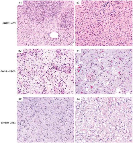
Intracranial mesenchymal tumors with FET-CREB fusions are a recently described group of neoplasms in children and young adults characterized by fusion of a FET family gene (usually EWSR1, but rarely FUS) to a CREB family transcription factor (ATF1, CREB1, or CREM), and have been variously termed intracranial angiomatoid fibrous histiocytoma or intracranial myxoid mesenchymal tumor. The clinical outcomes, histologic features, and genomic landscape are not well defined. Here, we studied 20 patients with intracranial mesenchymal tumors proven to harbor FET-CREB fusion by next-generation sequencing (NGS). The 16 female and four male patients had a median age of 14 years (range 4–70). Tumors were uniformly extra-axial or intraventricular and located at the cerebral convexities (n = 7), falx (2), lateral ventricles (4), tentorium (2), cerebellopontine angle (4), and spinal cord (1). NGS demonstrated that eight tumors harbored EWSR1-ATF1 fusion, seven had EWSR1-CREB1, four had EWSR1-CREM, and one had FUS-CREM. Tumors were uniformly well circumscribed and typically contrast enhancing with solid and cystic growth. Tumors with EWSR1-CREB1 fusions more often featured stellate/spindle cell morphology, mucin-rich stroma, and hemangioma-like vasculature compared to tumors with EWSR1-ATF1 fusions that most often featured sheets of epithelioid cells with mucin-poor collagenous stroma. These tumors demonstrated polyphenotypic immunoprofiles with frequent positivity for desmin, EMA, CD99, MUC4, and synaptophysin, but absence of SSTR2A, myogenin, and HMB45 expression. There was a propensity for local recurrence with a median progression-free survival of 12 months and a median overall survival of greater than 60 months, with three patients succumbing to disease (all with EWSR1-ATF1 fusions). In combination with prior case series, this study provides further insight into intracranial mesenchymal tumors with FET-CREB fusion, which represent a distinct group of CNS tumors encompassing both intracranial myxoid mesenchymal tumor and angiomatoid fibrous histiocytoma-like neoplasms.
中文翻译:

FET-CREB融合颅内间充质肿瘤——颅内粘液样间充质肿瘤和血管瘤样纤维组织细胞瘤样肿瘤谱的统一诊断
具有 FET-CREB 融合的颅内间充质肿瘤是最近描述的一组儿童和年轻成人肿瘤,其特征是 FET 家族基因(通常为EWSR1,但很少有FUS)与 CREB 家族转录因子(ATF1、CREB1或CREM),并被不同地称为颅内血管瘤样纤维组织细胞瘤或颅内粘液样间充质瘤。临床结果、组织学特征和基因组景观尚未明确定义。在这里,我们研究了 20 名颅内间充质肿瘤患者,通过下一代测序 (NGS) 证实具有 FET-CREB 融合。16 名女性和 4 名男性患者的中位年龄为 14 岁(范围 4-70)。肿瘤均位于轴外或脑室内,位于大脑凸面 (n = 7)、大脑镰 (2)、侧脑室 (4)、天幕 (2)、小脑桥脑角 (4) 和脊髓 (1)。NGS 证明 8 个肿瘤具有EWSR1 - ATF1融合,7 个具有EWSR1 - CREB1,4个具有EWSR1-CREM,一个有FUS - CREM。肿瘤界限清楚,通常对比增强,伴有实性和囊性生长。与具有EWSR1 - ATF1的肿瘤相比,具有EWSR1 - CREB1融合的肿瘤通常具有星状/梭形细胞形态、富含粘蛋白的基质和血管瘤样脉管系统融合最常见的特征是具有缺乏粘蛋白的胶原基质的上皮样细胞片。这些肿瘤表现出多表型免疫特征,结蛋白、EMA、CD99、MUC4 和突触素经常呈阳性,但缺乏 SSTR2A、肌细胞生成素和 HMB45 表达。有局部复发倾向,中位无进展生存期为 12 个月,中位总生存期大于 60 个月,其中 3 名患者死于疾病(均具有EWSR1 - ATF1融合)。结合之前的病例系列,本研究提供了对具有 FET-CREB 融合的颅内间充质肿瘤的进一步见解,它代表了一组独特的 CNS 肿瘤,包括颅内粘液样间充质肿瘤和血管瘤样纤维组织细胞瘤样肿瘤。
更新日期:2020-11-03
中文翻译:
FET-CREB融合颅内间充质肿瘤——颅内粘液样间充质肿瘤和血管瘤样纤维组织细胞瘤样肿瘤谱的统一诊断
具有 FET-CREB 融合的颅内间充质肿瘤是最近描述的一组儿童和年轻成人肿瘤,其特征是 FET 家族基因(通常为EWSR1,但很少有FUS)与 CREB 家族转录因子(ATF1、CREB1或CREM),并被不同地称为颅内血管瘤样纤维组织细胞瘤或颅内粘液样间充质瘤。临床结果、组织学特征和基因组景观尚未明确定义。在这里,我们研究了 20 名颅内间充质肿瘤患者,通过下一代测序 (NGS) 证实具有 FET-CREB 融合。16 名女性和 4 名男性患者的中位年龄为 14 岁(范围 4-70)。肿瘤均位于轴外或脑室内,位于大脑凸面 (n = 7)、大脑镰 (2)、侧脑室 (4)、天幕 (2)、小脑桥脑角 (4) 和脊髓 (1)。NGS 证明 8 个肿瘤具有EWSR1 - ATF1融合,7 个具有EWSR1 - CREB1,4个具有EWSR1-CREM,一个有FUS - CREM。肿瘤界限清楚,通常对比增强,伴有实性和囊性生长。与具有EWSR1 - ATF1的肿瘤相比,具有EWSR1 - CREB1融合的肿瘤通常具有星状/梭形细胞形态、富含粘蛋白的基质和血管瘤样脉管系统融合最常见的特征是具有缺乏粘蛋白的胶原基质的上皮样细胞片。这些肿瘤表现出多表型免疫特征,结蛋白、EMA、CD99、MUC4 和突触素经常呈阳性,但缺乏 SSTR2A、肌细胞生成素和 HMB45 表达。有局部复发倾向,中位无进展生存期为 12 个月,中位总生存期大于 60 个月,其中 3 名患者死于疾病(均具有EWSR1 - ATF1融合)。结合之前的病例系列,本研究提供了对具有 FET-CREB 融合的颅内间充质肿瘤的进一步见解,它代表了一组独特的 CNS 肿瘤,包括颅内粘液样间充质肿瘤和血管瘤样纤维组织细胞瘤样肿瘤。


 京公网安备 11010802027423号