Journal of Allergy and Clinical Immunology ( IF 14.2 ) Pub Date : 2020-10-23 , DOI: 10.1016/j.jaci.2020.09.009 Andreas Ronit 1 , Ronan M G Berg 2 , Jakob T Bay 3 , Anna K Haugaard 3 , Magnus G Ahlström 4 , Kristoffer S Burgdorf 3 , Henrik Ullum 3 , Sara B Rørvig 5 , Klaus Tjelle 6 , Nicolai B Foss 6 , Thomas Benfield 1 , Hanne Vibeke Marquart 3 , Ronni R Plovsing 6
|
Background
Severe immunopathology may drive the deleterious manifestations that are observed in the advanced stages of coronavirus disease 2019 (COVID-19) but are poorly understood.
Objective
Our aim was to phenotype leukocyte subpopulations and the cytokine milieu in the lungs and blood of critically ill patients with COVID-19 acute respiratory distress syndrome (ARDS).
Methods
We consecutively included patients less than 72 hours after intubation following informed consent from their next of kin. Bronchoalveolar lavage fluid was evaluated by microscopy; bronchoalveolar lavage fluid and blood were assessed by 10-color flow cytometry and a multiplex cytokine panel.
Results
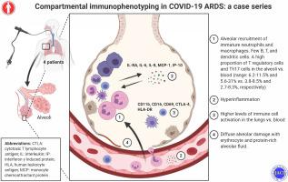
Four mechanically ventilated patients (aged 40-75 years) with moderate-to-severe COVID-19 ARDS were included. Immature neutrophils dominated in both blood and lungs, whereas CD4 and CD8 T-cell lymphopenia was observed in the 2 compartments. However, regulatory T cells and TH17 cells were found in higher fractions in the lung. Lung CD4 and CD8 T cells and macrophages expressed an even higher upregulation of activation markers than in blood. A wide range of cytokines were expressed at high levels both in the blood and in the lungs, most notably, IL-1RA, IL-6, IL-8, IP-10, and monocyte chemoattactant protein-1, consistent with hyperinflammation.
Conclusion
COVID-19 ARDS exhibits a distinct immunologic profile in the lungs, with a depleted and exhausted CD4 and CD8 T-cell population that resides within a heavily hyperinflammatory milieu.
中文翻译:
COVID-19 ARDS 中的隔室免疫表型分析:病例系列
背景
严重的免疫病理学可能会导致在 2019 年冠状病毒病 (COVID-19) 的晚期阶段观察到的有害表现,但人们对此知之甚少。
客观的
我们的目的是对 COVID-19 急性呼吸窘迫综合征 (ARDS) 重症患者的肺和血液中的白细胞亚群和细胞因子环境进行表型分析。
方法
在获得直系亲属的知情同意后,我们连续纳入插管后不到 72 小时的患者。通过显微镜评估支气管肺泡灌洗液;通过 10 色流式细胞术和多重细胞因子面板评估支气管肺泡灌洗液和血液。
结果
包括四名中度至重度 COVID-19 ARDS 的机械通气患者(年龄 40-75 岁)。未成熟的中性粒细胞在血液和肺中占主导地位,而在 2 个隔室中观察到 CD4 和 CD8 T 细胞淋巴细胞减少。然而,在肺的较高部分中发现了调节性 T 细胞和 T H 17 细胞。与血液相比,肺 CD4 和 CD8 T 细胞和巨噬细胞表达的活化标志物上调甚至更高。多种细胞因子在血液和肺中均以高水平表达,最显着的是 IL-1RA、IL-6、IL-8、IP-10 和单核细胞趋化蛋白 1,与过度炎症一致。
结论
COVID-19 ARDS 在肺部表现出独特的免疫学特征,其 CD4 和 CD8 T 细胞群处于极度高炎症环境中。


 京公网安备 11010802027423号