IRBM ( IF 4.8 ) Pub Date : 2020-10-28 , DOI: 10.1016/j.irbm.2020.10.007 Y. Zhang , J. Duan , Y. Sa , Y. Guo
|
Background
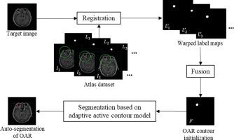
Accurate delineation of organs at risk (OARs) is critical in radiotherapy. Manual delineation is tedious and suffers from both interobserver and intraobserver variability. Automatic segmentation of brain MR images has a wide range of applications in brain tumor radiotherapy. In this paper, we propose a multi-atlas based adaptive active contour model for OAR automatic segmentation in brain MR images.
Methods
The proposed method consists of two parts: multi-atlas based OAR contour initiation and an adaptive edge and local region based active contour evolution. In the adaptive active contour model, we define an energy functional with an adaptive edge intensity fitting force which is responsible for evaluating contour inwards or outwards, and a local region intensity fitting force which guides the evolution of the contour.
Results
Experimental results show that the proposed method achieved more accurate segmentation results in brainstem, eyes and lens automatic segmentation with the Dice Similar Coefficient (DSC) value of 87.19%, 91.96%, 77.11% respectively. Besides, the dosimetric parameters also demonstrate the high consistency of the manual OAR delineations and the auto segmentation results of the proposed method in brain tumor radiotherapy.
Conclusions
The geometric and dosimetric evaluations show the desirable performance of the proposed method on the application of OARs segmentations in brain tumor radiotherapy.
中文翻译:
基于多图集的自适应主动轮廓模型及其在脑 MR 图像中风险器官分割的应用
背景
准确描绘危险器官 (OAR) 在放射治疗中至关重要。手动描绘是乏味的,并且受到观察者间和观察者内变化的影响。脑部MR图像的自动分割在脑肿瘤放射治疗中有着广泛的应用。在本文中,我们提出了一种基于多图集的自适应主动轮廓模型,用于脑 MR 图像中的 OAR 自动分割。
方法
所提出的方法由两部分组成:基于多图集的 OAR 轮廓起始和基于自适应边缘和局部区域的主动轮廓演化。在自适应主动轮廓模型中,我们定义了一个能量泛函,具有自适应边缘强度拟合力,负责评估轮廓向内或向外,以及指导轮廓演化的局部区域强度拟合力。
结果
实验结果表明,该方法在脑干、眼睛和晶状体自动分割中取得了更准确的分割结果,Dice相似系数(DSC)值分别为87.19%、91.96%、77.11%。此外,剂量学参数还证明了手动 OAR 描绘和所提出方法在脑肿瘤放射治疗中的自动分割结果的高度一致性。
结论
几何和剂量学评估显示了所提出的方法在脑肿瘤放射治疗中应用 OAR 分割的理想性能。


 京公网安备 11010802027423号