Neurosurgical Review ( IF 2.8 ) Pub Date : 2020-10-24 , DOI: 10.1007/s10143-020-01405-0 Helbert de Oliveira Manduca Palmiero 1 , Davi Jorge Fontoura Solla 1 , Leonardo Borges Dos Santos 1 , Manoel Jacobsen Teixeira 1 , Eberval Gadelha Figueiredo 1
|
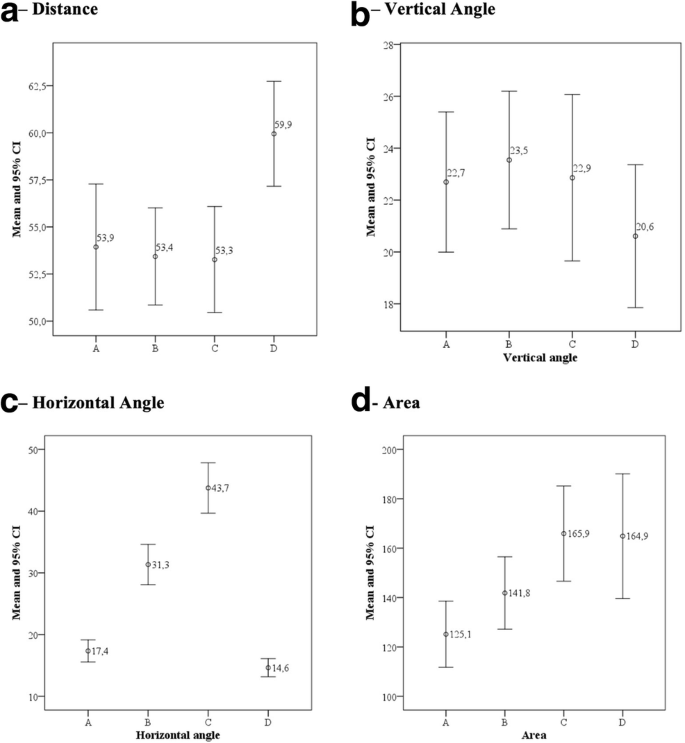
The posteroinferior region of the thalamus is formed by the pulvinar, and it is surgically accessed through the infratentorial supracerebellar approach, between the midline and the retromastoid region. This study aimed to compare the paramedian, lateral, extreme lateral, and contralateral paramedian corridors with the posteroinferior thalamus through a suboccipital craniotomy and an infratentorial supracerebellar access. Ten cadavers were studied, and the microsurgical dissections were accompanied by the measurement of the variables using a neuronavigation system. Statistical analysis was performed using analysis of variance (ANOVA). The distance between the access midpoint at the cranial surface and pulvinar varied between 53.3 and 53.9 mm, the contralateral access being an exception (59.9 mm). The vertical angle ranged from 20.6° in the contralateral access to 23.5° in the lateral access. There was a gradual increase in the horizontal angle between the paramedian (17.4°), lateral (31.3°), and extreme lateral (43.7°) accesses. But, this angle in the contralateral access was 14.6°, similar to that of the paramedian access. The exposed area of the thalamus was 125.1 mm2 in the paramedian access, 141.8 mm2 in the lateral access, and 165.9 mm2 in the extreme lateral access, which was similar to that of the contralateral access (164.9 mm2). The horizontal view angle increased with lateralization of the access, which facilitated microscopic visualization. With regard to the exposure of the microsurgical anatomy, the extreme lateral and contralateral accesses circumvent the neural and vascular obstacles at the midline, allowing a larger area of anatomical exposure.
Graphical abstract
中文翻译:
后下丘脑幕下小脑上入路变体的解剖学、定性和定量评估
丘脑后下区由丘脑形成,可通过幕下小脑上入路手术进入,位于中线和乳突后区之间。本研究旨在通过枕下开颅手术和幕下小脑上通路比较旁正中、外侧、极外侧和对侧旁正中通道与后下丘脑。研究了十具尸体,显微外科解剖伴随着使用神经导航系统测量变量。使用方差分析(ANOVA)进行统计分析。颅面入路中点和骨盆之间的距离在 53.3 和 53.9 毫米之间变化,对侧入路是一个例外(59.9 毫米)。垂直角度范围为 20。对侧通路 6° 到侧通路 23.5°。旁正中(17.4°)、外侧(31.3°)和极外侧(43.7°)通路之间的水平角逐渐增加。但是,对侧入路的这个角度为 14.6°,与旁正中入路的角度相似。丘脑的暴露面积为 125.1 mm2在正中访问,141.8毫米2在横向接入,165.9毫米2在极外侧访问,这是类似于对侧访问(164.9毫米2)。水平视角随着通路的侧化而增加,这有利于微观可视化。在显微解剖暴露方面,最外侧和对侧通路绕过中线的神经和血管障碍,允许更大面积的解剖暴露。


 京公网安备 11010802027423号