当前位置:
X-MOL 学术
›
J. Am. Chem. Soc.
›
论文详情
Our official English website, www.x-mol.net, welcomes your feedback! (Note: you will need to create a separate account there.)
Probing Nanoscale Diffusional Heterogeneities in Cellular Membranes through Multidimensional Single-Molecule and Super-Resolution Microscopy
Journal of the American Chemical Society ( IF 15.0 ) Pub Date : 2020-10-21 , DOI: 10.1021/jacs.0c08426 Rui Yan 1, 2 , Kun Chen 1, 2 , Ke Xu 1, 2
Journal of the American Chemical Society ( IF 15.0 ) Pub Date : 2020-10-21 , DOI: 10.1021/jacs.0c08426 Rui Yan 1, 2 , Kun Chen 1, 2 , Ke Xu 1, 2
Affiliation
|
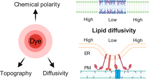
Diffusion properties notably determine the behavior of biomembranes. Here we report the concurrent nanoscale fine-mapping of membrane topography, diffusivity, and packing order in live mammalian cells through a synergy of single-molecule and super-resolution methods. By identifying a bright, lipophilic fluorescence turn-on probe that enables sustained single-molecule imaging of cellular membranes under stroboscopic excitation, we accumulate the positions and transient displacements of >106 probe molecules to achieve super-resolution topography and diffusivity mapping. We thus determine a trend that the membrane diffusivity drops with increased lipid packing order when comparing the endoplasmic reticulum (ER) membrane, plasma membrane, and nanodomains induced by cholera toxin B. Utilizing our nanoscale mapping capability, we further unveil reduced diffusivity in the ER membrane at ER-plasma membrane contact sites. By next integrating spectrally resolved single-molecule imaging, we show that this localized diffusion slowdown is not due to altered lipid packing order but may instead be attributed to local protein crowding. Our integrated multidimensional single-molecule approach thus unveils and differentiates between nanoscale diffusional heterogeneities of different origins in live-cell membranes.
中文翻译:

通过多维单分子和超分辨率显微镜探测细胞膜中的纳米级扩散异质性
扩散特性显着地决定了生物膜的行为。在这里,我们通过单分子和超分辨率方法的协同作用报告了活哺乳动物细胞中膜形貌、扩散率和堆积顺序的并行纳米级精细映射。通过识别一种明亮的亲脂荧光开启探针,该探针能够在频闪激发下对细胞膜进行持续的单分子成像,我们积累了 >106 个探针分子的位置和瞬态位移,以实现超分辨率地形和扩散映射。因此,当比较霍乱毒素 B 诱导的内质网 (ER) 膜、质膜和纳米域时,我们确定膜扩散率随着脂质堆积顺序的增加而下降。利用我们的纳米级映射能力,我们进一步揭示了 ER 膜接触部位的 ER 膜扩散率降低。通过接下来整合光谱分辨单分子成像,我们表明这种局部扩散放缓不是由于脂质堆积顺序改变,而是可能归因于局部蛋白质拥挤。因此,我们集成的多维单分子方法揭示并区分了活细胞膜中不同来源的纳米级扩散异质性。
更新日期:2020-10-21
中文翻译:
通过多维单分子和超分辨率显微镜探测细胞膜中的纳米级扩散异质性
扩散特性显着地决定了生物膜的行为。在这里,我们通过单分子和超分辨率方法的协同作用报告了活哺乳动物细胞中膜形貌、扩散率和堆积顺序的并行纳米级精细映射。通过识别一种明亮的亲脂荧光开启探针,该探针能够在频闪激发下对细胞膜进行持续的单分子成像,我们积累了 >106 个探针分子的位置和瞬态位移,以实现超分辨率地形和扩散映射。因此,当比较霍乱毒素 B 诱导的内质网 (ER) 膜、质膜和纳米域时,我们确定膜扩散率随着脂质堆积顺序的增加而下降。利用我们的纳米级映射能力,我们进一步揭示了 ER 膜接触部位的 ER 膜扩散率降低。通过接下来整合光谱分辨单分子成像,我们表明这种局部扩散放缓不是由于脂质堆积顺序改变,而是可能归因于局部蛋白质拥挤。因此,我们集成的多维单分子方法揭示并区分了活细胞膜中不同来源的纳米级扩散异质性。


 京公网安备 11010802027423号