Journal of Fluorescence ( IF 2.7 ) Pub Date : 2020-10-19 , DOI: 10.1007/s10895-020-02637-5 Vladimir Kanygin 1 , Alexander Zaboronok 1, 2, 3 , Iuliia Taskaeva 4, 5, 6 , Evgenii Zavjalov 1, 7 , Rinat Mukhamadiyarov 1, 8 , Aleksandr Kichigin 1 , Anna Kasatova 1, 6 , Ivan Razumov 1, 7 , Roman Sibirtsev 1 , Bryan J Mathis 2
|
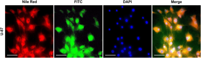
Boron neutron capture therapy (BNCT), a binary cancer therapeutic modality, has moved to a new phase since development of accelerator-based neutron sources and establishment of BNCT centers in Finland and Japan. That stimulated efforts for better boron delivery agent development. As liposomes have shown effective boron delivery properties and sufficient tumor retention, fluorescent liposome labelling may serve as a rapid method to study initial ability of newly synthesized liposomes to be captured by tumor cells prior to experiments on boron accumulation and neutron irradiation. In this work, we studied the accumulation and biodistribution of pegylated liposomes with encapsulated borocaptate (BSH) and a fluorescent label (Nile Red) in U87 (human glioblastoma), SW-620 (human colon carcinoma), SK-MEL-28 (human melanoma), FetMSC (mesenchymal human embryo stem cells), and EMBR (primary embryocytes) cell lines as well as an orthotopic xenograft model of U87 glioma in SCID mice. Results indicate that fluorescent microscopy is effective at determining the intracellular localization of the liposomes using a fluorescent label. The synthesized, pegylated liposomes showed higher accumulation in tumors compared to normal cells, with characteristic concentration peaks in SW-620 and U87 cell lines, and provided in vivo tumor selectivity with several-fold higher tumor tissue fluorescence at the 6-h timepoint.
中文翻译:
荧光标记的含硼酸酯脂质体的体外和体内评价
自开发基于加速器的中子源并在芬兰和日本建立 BNCT 中心以来,硼中子俘获疗法 (BNCT) 是一种二元癌症治疗方式,已进入新阶段。这刺激了更好的硼输送剂开发的努力。由于脂质体已显示出有效的硼递送特性和足够的肿瘤保留,荧光脂质体标记可作为一种快速方法来研究新合成的脂质体在硼积累和中子辐照实验之前被肿瘤细胞捕获的初始能力。在这项工作中,我们研究了聚乙二醇化脂质体在 U87(人胶质母细胞瘤)、SW-620(人结肠癌)、SK-MEL-28(人黑色素瘤),FetMSC(间充质人胚胎干细胞)和 EMBR(原代胚胎细胞)细胞系以及 SCID 小鼠中 U87 神经胶质瘤的原位异种移植模型。结果表明荧光显微镜在使用荧光标记确定脂质体的细胞内定位方面是有效的。与正常细胞相比,合成的聚乙二醇化脂质体在肿瘤中显示出更高的积累,在 SW-620 和 U87 细胞系中具有特征浓度峰,并在 6 小时时间点提供了体内肿瘤选择性高几倍的肿瘤组织荧光。结果表明荧光显微镜在使用荧光标记确定脂质体的细胞内定位方面是有效的。与正常细胞相比,合成的聚乙二醇化脂质体在肿瘤中显示出更高的积累,在 SW-620 和 U87 细胞系中具有特征浓度峰,并在 6 小时时间点提供了体内肿瘤选择性高几倍的肿瘤组织荧光。结果表明荧光显微镜在使用荧光标记确定脂质体的细胞内定位方面是有效的。与正常细胞相比,合成的聚乙二醇化脂质体在肿瘤中显示出更高的积累,在 SW-620 和 U87 细胞系中具有特征浓度峰,并在 6 小时时间点提供了体内肿瘤选择性高几倍的肿瘤组织荧光。


 京公网安备 11010802027423号