Biomaterials Advances ( IF 7.9 ) Pub Date : 2020-10-17 , DOI: 10.1016/j.msec.2020.111639 K. Zafeiris , D. Brasinika , A. Karatza , Elias Koumoulos , I.K. Karoussis , K. Kyriakidou , C.A. Charitidis
|
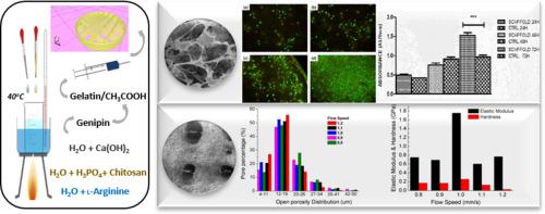
Additive manufacturing holds promise for the fabrication of three-dimensional scaffolds with precise geometry, to serve as substrates for the guided regeneration of natural tissue. In this work, a bioinspired approach is adopted for the synthesis of hybrid hydroxyapatite hydrogels, which were subsequently printed to form 3D scaffolds for bone tissue engineering applications. These hydrogels consist of hydroxyapatite nanocrystals, biomimetically synthesized in the presence of both chitosan and l-arginine. To improve their mechanical properties, chemical crosslinking was performed using a natural crosslinking agent (genipin), and their rheology was modified by employing an acetic acid/gelatin solution. Regarding the 3D printing process, several parameters (flow, infill and perimeter speed) were studied in order to accurately produce scaffolds with predesigned geometry and micro-architecture, while also applying low printing temperature (15 °C). Following the printing procedure, the 3D scaffolds were freeze dried in order to remove the entrapped solvents and therefore, obtain a porous interconnected network. Evaluation of porosity was performed using micro-computed tomography and nanomechanical properties were assessed through nanoindentation. Results of both characterization techniques, showed that the scaffolds' porosity as well as their modulus values, fall within the corresponding range of the respective values of cancellous bone. The biocompatibility of the 3D printed scaffolds was assessed using MG63 human osteosarcoma cells for 7 days of culturing. Cell viability was evaluated by MTT assay as well as double staining and visualized under fluorescence microscopy, while cell morphology was analyzed through scanning electron microscopy. Biocompatibility tests, revealed that the scaffolds constitute a cell-friendly environment, allowed them to adhere on the scaffolds' surface, increase their population and maintain high levels of viability.
中文翻译:
用于骨组织工程应用的羟基磷灰石-壳聚糖-genipin复合支架的增材制造
增材制造有望制造出具有精确几何形状的三维支架,以用作自然组织的引导再生的基质。在这项工作中,采用了生物启发的方法来合成混合羟基磷灰石水凝胶,随后将其印刷形成用于骨组织工程应用的3D支架。这些水凝胶由羟基磷灰石纳米晶体的,在脱乙酰壳多糖的存在和仿生合成升-精氨酸 为了改善它们的机械性能,使用天然交联剂(genipin)进行化学交联,并通过使用乙酸/明胶溶液改变其流变性。关于3D打印过程,研究了几个参数(流动,填充和周长速度),以便精确地生产具有预先设计的几何形状和微结构的支架,同时还适用较低的打印温度(15°C)。在印刷程序之后,将3D支架冷冻干燥,以去除残留的溶剂,因此获得了多孔的互连网络。使用微计算机断层摄影术进行孔隙率评估,并通过纳米压痕评估纳米机械性能。两种表征技术的结果表明,脚手架 孔隙率及其模量值落入松质骨各自值的相应范围内。使用MG63人骨肉瘤细胞培养7天,评估3D打印支架的生物相容性。通过MTT测定以及双重染色评价细胞活力,并在荧光显微镜下可视化,同时通过扫描电子显微镜分析细胞形态。生物相容性测试表明,该支架构成了一个对细胞友好的环境,使它们能够粘附在支架表面上,增加了其种群并维持了高水平的生存能力。使用MG63人骨肉瘤细胞培养7天,评估3D打印支架的生物相容性。通过MTT测定以及双重染色评价细胞活力,并在荧光显微镜下可视化,同时通过扫描电子显微镜分析细胞形态。生物相容性测试表明,该支架构成了一个对细胞友好的环境,使它们能够粘附在支架表面上,增加了其种群并维持了高水平的生存能力。使用MG63人骨肉瘤细胞培养7天,评估3D打印支架的生物相容性。通过MTT测定以及双重染色评价细胞活力,并在荧光显微镜下可视化,同时通过扫描电子显微镜分析细胞形态。生物相容性测试表明,该支架构成了一个对细胞友好的环境,使它们能够粘附在支架表面上,增加了种群并维持了高水平的生存能力。


 京公网安备 11010802027423号