当前位置:
X-MOL 学术
›
Brain Pathol.
›
论文详情
Our official English website, www.x-mol.net, welcomes your feedback! (Note: you will need to create a separate account there.)
The use and limitations of single‐cell mass cytometry for studying human microglia function
Brain Pathology ( IF 6.4 ) Pub Date : 2020-10-15 , DOI: 10.1111/bpa.12909 Camila Fernández-Zapata 1 , Julia K H Leman 1 , Josef Priller 1, 2, 3 , Chotima Böttcher 1
Brain Pathology ( IF 6.4 ) Pub Date : 2020-10-15 , DOI: 10.1111/bpa.12909 Camila Fernández-Zapata 1 , Julia K H Leman 1 , Josef Priller 1, 2, 3 , Chotima Böttcher 1
Affiliation
|
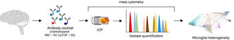
Microglia, the resident innate immune cells of the central nervous system (CNS), play an important role in brain development and homoeostasis, as well as in neuroinflammatory, neurodegenerative and psychiatric diseases. Studies in animal models have been used to determine the origin and development of microglia, and how these cells alter their transcriptional and phenotypic signatures during CNS pathology. However, little is known about their human counterparts. Recent studies in human brain samples have harnessed the power of multiplexed single‐cell technologies such as single‐cell RNA sequencing (scRNA‐seq) and mass cytometry (cytometry by time‐of‐flight [CyTOF]) to provide a comprehensive molecular view of human microglia in healthy and diseased brains. CyTOF is a powerful tool to study high‐dimensional protein expression of human microglia (huMG) at the single‐cell level. This technology widens the possibilities of high‐throughput quantification (of over 60 targeted molecules) at a single‐cell resolution. CyTOF can be combined with scRNA‐seq for comprehensive analysis, as it allows single‐cell analysis of post‐translational modifications of proteins, which provides insights into cell signalling dynamics in targeted cells. In addition, imaging mass cytometry (IMC) has recently become commercially available, and will be useful for analysing multiple cell types in human brain sections. IMC leverages mass spectrometry to acquire spatial data of cell–cell interactions on tissue sections, using (theoretically) over 40 markers at the same time. In this review, we summarise recent studies of huMG using CyTOF and IMC analyses. The uses and limitations as well as future directions of these technologies are discussed.
中文翻译:

单细胞质谱在研究人类小胶质细胞功能中的应用和局限性
小胶质细胞是中枢神经系统 (CNS) 的固有先天免疫细胞,在大脑发育和体内平衡以及神经炎症、神经退行性疾病和精神疾病中发挥重要作用。动物模型研究已被用于确定小胶质细胞的起源和发育,以及这些细胞在 CNS 病理学过程中如何改变其转录和表型特征。然而,人们对它们的人类对应物知之甚少。最近对人脑样本的研究利用了多重单细胞技术的力量,例如单细胞 RNA 测序 (scRNA-seq) 和质谱流式细胞术(飞行时间流式细胞术 [CyTOF]),以提供全面的分子视角。健康和患病大脑中的人类小胶质细胞。CyTOF 是在单细胞水平研究人类小胶质细胞 (huMG) 高维蛋白表达的强大工具。该技术扩大了以单细胞分辨率进行高通量定量(超过 60 个目标分子)的可能性。CyTOF 可以与 scRNA-seq 结合进行综合分析,因为它允许对蛋白质的翻译后修饰进行单细胞分析,从而深入了解靶细胞中的细胞信号动力学。此外,成像质量流式细胞术 (IMC) 最近已商业化,可用于分析人脑切片中的多种细胞类型。IMC 利用质谱法来获取组织切片上细胞间相互作用的空间数据,同时使用(理论上)超过 40 个标记。在这次审查中,我们总结了最近使用 CyTOF 和 IMC 分析对 huMG 的研究。讨论了这些技术的用途和局限性以及未来的方向。
更新日期:2020-12-17
中文翻译:
单细胞质谱在研究人类小胶质细胞功能中的应用和局限性
小胶质细胞是中枢神经系统 (CNS) 的固有先天免疫细胞,在大脑发育和体内平衡以及神经炎症、神经退行性疾病和精神疾病中发挥重要作用。动物模型研究已被用于确定小胶质细胞的起源和发育,以及这些细胞在 CNS 病理学过程中如何改变其转录和表型特征。然而,人们对它们的人类对应物知之甚少。最近对人脑样本的研究利用了多重单细胞技术的力量,例如单细胞 RNA 测序 (scRNA-seq) 和质谱流式细胞术(飞行时间流式细胞术 [CyTOF]),以提供全面的分子视角。健康和患病大脑中的人类小胶质细胞。CyTOF 是在单细胞水平研究人类小胶质细胞 (huMG) 高维蛋白表达的强大工具。该技术扩大了以单细胞分辨率进行高通量定量(超过 60 个目标分子)的可能性。CyTOF 可以与 scRNA-seq 结合进行综合分析,因为它允许对蛋白质的翻译后修饰进行单细胞分析,从而深入了解靶细胞中的细胞信号动力学。此外,成像质量流式细胞术 (IMC) 最近已商业化,可用于分析人脑切片中的多种细胞类型。IMC 利用质谱法来获取组织切片上细胞间相互作用的空间数据,同时使用(理论上)超过 40 个标记。在这次审查中,我们总结了最近使用 CyTOF 和 IMC 分析对 huMG 的研究。讨论了这些技术的用途和局限性以及未来的方向。


 京公网安备 11010802027423号