Journal of Materials Science: Materials in Medicine ( IF 3.7 ) Pub Date : 2020-10-14 , DOI: 10.1007/s10856-020-06446-x A M Porras Hernández 1 , H Pohlit 1 , F Sjögren 1 , L Shi 2 , D Ossipov 2, 3 , M Antfolk 4, 5 , M Tenje 1
|
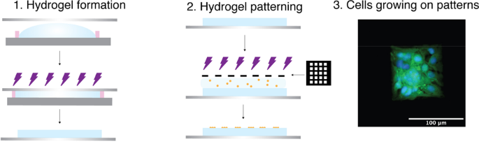
In this work, we present a method to fabricate a hyaluronic acid (HA) hydrogel with spatially controlled cell-adhesion properties based on photo-polymerisation cross-linking and functionalization. The approach utilises the same reaction pathway for both steps meaning that it is user-friendly and allows for adaptation at any stage during the fabrication process. Moreover, the process does not require any additional cross-linkers. The hydrogel is formed by UV-initiated radical addition reaction between acrylamide (Am) groups on the HA backbone. Cell adhesion is modulated by functionalising the adhesion peptide sequence arginine–glycine–aspartate onto the hydrogel surface via radical mediated thiol–ene reaction using the non-reacted Am groups. We show that 10 × 10 µm2 squares could be patterned with sharp features and a good resolution. The smallest area that could be patterned resulting in good cell adhesion was 25 × 25 µm2 squares, showing single-cell adhesion. Mouse brain endothelial cells adhered and remained in culture for up to 7 days on 100 × 100 µm2 square patterns. We see potential for this material combination for future use in novel organ-on-chip models and tissue engineering where the location of the cells is of importance and to further study endothelial cell biology.
中文翻译:
通过直接紫外线介导的 RGD 连接控制细胞粘附在生物衍生的体外细胞培养支架上的简化方法
在这项工作中,我们提出了一种基于光聚合交联和功能化制备具有空间控制细胞粘附特性的透明质酸 (HA) 水凝胶的方法。该方法对两个步骤使用相同的反应途径,这意味着它是用户友好的,并允许在制造过程中的任何阶段进行调整。此外,该过程不需要任何额外的交联剂。水凝胶是通过紫外线引发的 HA 主链上丙烯酰胺 (Am) 基团之间的自由基加成反应形成的。细胞粘附是通过使用未反应的 Am 基团通过自由基介导的硫醇 - 烯反应将粘附肽序列精氨酸 - 甘氨酸 - 天冬氨酸官能化到水凝胶表面来调节的。我们表明 10 × 10 µm 2正方形可以用清晰的特征和良好的分辨率进行图案化。可形成良好细胞粘附的图案化最小面积为 25 × 25 µm 2方格,显示单细胞粘附。小鼠脑内皮细胞在 100 × 100 µm 2方形图案上粘附并保持培养长达 7 天。我们看到这种材料组合在未来用于新型器官芯片模型和组织工程中的潜力,其中细胞的位置很重要,并进一步研究内皮细胞生物学。


 京公网安备 11010802027423号