Micron ( IF 2.4 ) Pub Date : 2020-10-13 , DOI: 10.1016/j.micron.2020.102963 Atharva Damle , Sangeetha Muthusamy , Reetoja Nag , Raunak Kumar Das , Priyanka Srivastava
|
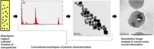
The study employs conventional techniques and quantitative image analysis tools to characterize alginate-capped nanosilver synthesized by green methods. Sodium Alginate (0.5 %, 1 % and 2 %) was used as a reducing and stabilizing agent. Presence of particles was confirmed by UV–vis Spectroscopy, with absorbance maxima of 412−413 nm for 0.5 %, 1 % and 2 % of polymer. Hydrodynamic sizes of particles recorded for 0.5 %, 1 % and 2 % polymer were 128.4 ± 1.5, 129.9 ± 3.6 and 148.6 ± 1.0 nm by DLS. TEM revealed roughly spherical to cuboidal particles ranging from 15−20 nm and clusters of 100 nm and Energy Dispersive X-ray Spectroscopy confirmed the presence of silver in the particles. Analysis of the TEM images was done in MATLAB R2016b using histogram equalisation for image enhancement and entropy filtering for image segmentation. These techniques revealed the surface pores and polymer distribution around the particle. Statistical analysis (ANOVA) was performed for the measured fractal dimensions of nanoparticles with polymer coating, width of particle together with polymer coating, and thickness of only polymer coating around the particle for various study groups. Significant differences (p < 0.05) were found both between and within the study groups for fractal dimensions of nanoparticles with polymer coating, width of nanoparticles and thickness of polymer coating alone. The analysis was successful in confirming presence and thickness of polymer layer on particles.
中文翻译:
海藻酸盐封端的纳米银的透射电子显微照片的分形和熵分析
该研究采用常规技术和定量图像分析工具来表征通过绿色方法合成的藻酸盐封端的纳米银。海藻酸钠(0.5%,1%和2%)用作还原剂和稳定剂。紫外可见光谱法证实了颗粒的存在,对于0.5%,1%和2%的聚合物,最大吸收值为412-413 nm。通过DLS记录的0.5%,1%和2%聚合物的颗粒的流体力学尺寸为128.4±1.5、129.9±3.6和148.6±1.0nm。透射电镜显示大约15-20 nm的球形到长方体颗粒和100 nm的簇,并且能量色散X射线光谱证实颗粒中存在银。TEM图像的分析是在MATLAB R2016b中使用直方图均衡进行图像增强,并使用熵过滤进行图像分割。这些技术揭示了颗粒周围的表面孔隙和聚合物分布。进行统计分析(ANOVA),以测量各个研究组具有聚合物涂层的纳米颗粒的分形维数,颗粒与聚合物涂层的宽度以及仅颗粒周围的聚合物涂层的厚度。重大差异(在研究组之间和研究组内和内部都发现了具有聚合物涂层的纳米颗粒的分形尺寸,纳米颗粒的宽度和单独的聚合物涂层的厚度(p <0.05)。该分析成功地证实了颗粒上聚合物层的存在和厚度。


 京公网安备 11010802027423号