Applied Microbiology and Biotechnology ( IF 5 ) Pub Date : 2020-10-12 , DOI: 10.1007/s00253-020-10952-x Mengxiao Wang , Jingxuan Ma , Xuewei Wang , Zhijun Wang , Lingyi Tang , Haoming Chen , Zhen Li
|
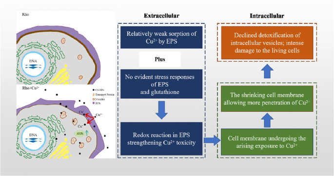
The red yeast (Rhodotorula mucilaginosa: Rho) has abundant extracellular polymeric substances (EPS) and intracellular vesicles (Ves). This study explored the mechanisms of Rho to resist Cu toxicity from extracellular to intracellular, i.e., EPS, membrane, and Ves. The Cu2+ concentrations were set from 0 to 200 mg/L. In contrast to other heavy metals (e.g., Pb2+), low Cu2+ stress has no evident stimulation to EPS production. In particular, GSH content in EPS did not show significant changes. The Cu removal was decreased from ~ 35 to ~ 0% as Cu stress raised from 0 to 200 mg/L, which confirmed the low binding of Cu cations to EPS. Moreover, redox peaks at − 0.35 V (reduction) and − 0.02 V (oxidation) in EPS were observed based on electrochemical analysis. Subsequently, the potential Haber-Weiss reaction in EPS lowered fungal ability to shield against the Cu toxicity. Then, the contrast of Cu concentration between the extracellular and intracellular regions was enlarged. Moreover, the thickness of cell membrane decreased from 450 to 116 nm during the elevation of Cu stress. These accelerated the transport of Cu cations into intracellular, but the redox reaction in both cell membrane and intracellular region was limited. Under transmission electron microscopy, the intracellular Ves showed evident sorption of Cu cations (100 mg/L). However, the Ves started to deform and gradually lost their activity at 200 mg/L. Therefore, this study successfully elucidated the correlated extracellular and intracellular mechanisms of metal detoxification by yeast.
Key points
•This study provides a comprehensive explanation for the invasion of Cu 2+ into fungal (Rhodotorula mucilaginosa) cells based on microbial physiological and biochemical analysis, electrochemical analysis, and transmitted electron microscopy.
•Cu nanoparticles are involved in redox reactions in the EPS, thus greatly reducing the prophase protection for fungal cells by EPS.
•At 200 mg/L Cu 2+ stress, deformation of cell membrane intensifies the contrast of Cu concentrations between extra- and intracellular regions. This further suppresses the transportation of Cu 2+ by intracellular vesicles.
中文翻译:
红酵母Rhodotorula mucilaginosa对Cu(II)的解毒作用:从细胞外到细胞内
红色酵母(Rhodotorula mucilaginosa:Rho)具有丰富的细胞外聚合物(EPS)和细胞内囊泡(Ves)。这项研究探索了Rho抵抗铜从细胞外到细胞内毒性的机制,即EPS,膜和Ves。Cu 2+浓度设定为0至200mg / L。与其他重金属(例如Pb 2+)相比,Cu 2+低压力没有明显刺激EPS的产生。特别是,EPS中的GSH含量没有显示出明显变化。随着铜应力从0升高到200 mg / L,铜的去除率从〜35降低到〜0%,这证实了铜阳离子与EPS的低结合力。此外,基于电化学分析,在EPS中观察到氧化还原峰在-0.35 V(还原)和-0.02 V(氧化)。随后,EPS中潜在的Haber-Weiss反应降低了真菌抵抗铜毒性的能力。然后,细胞外和细胞内区域之间的铜浓度的对比扩大了。此外,在铜应力升高期间,细胞膜的厚度从450纳米降低到116纳米。这些促进了铜阳离子向细胞内的转运,但是在细胞膜和细胞内区域的氧化还原反应受到限制。在透射电镜下,细胞内的Ves显示出明显的Cu阳离子吸附(100mg / L)。但是,Ves开始变形,并在200 mg / L时逐渐失去活性。因此,这项研究成功阐明了酵母对金属进行解毒的相关细胞外和细胞内机制。
关键点
•本研究 基于微生物生理生化分析,电化学分析和透射电子显微镜,为Cu 2+ 侵入真菌(Rhodotorula mucilaginosa)细胞提供了全面的解释。
•铜纳米粒子参与EPS中的氧化还原反应,因此大大降低了EPS对真菌细胞的前期保护。
•在200 mg / L的Cu 2+ 胁迫下,细胞膜的变形会增强细胞外和细胞内区域之间Cu浓度的对比度。这进一步抑制了 细胞内小泡对Cu 2+ 的转运。


 京公网安备 11010802027423号