当前位置:
X-MOL 学术
›
Chem. Sci.
›
论文详情
Our official English website, www.x-mol.net, welcomes your feedback! (Note: you will need to create a separate account there.)
LDL-mimetic lipid nanoparticles prepared by surface KAT ligation for in vivo MRI of atherosclerosis
Chemical Science ( IF 8.4 ) Pub Date : 2020-10-07 , DOI: 10.1039/d0sc04106h Alessandro Fracassi 1, 2, 3, 4 , Jianbo Cao 5, 6, 7, 8, 9 , Naoko Yoshizawa-Sugata 10, 11, 12, 13 , Éva Tóth 14, 15, 16, 17, 18 , Corey Archer 2, 4, 19, 20 , Olivier Gröninger 4, 21, 22, 23 , Emanuela Ricciotti 7, 8, 9, 24 , Soon Yew Tang 7, 8, 9, 24 , Stephan Handschin 2, 4, 25, 26 , Jean-Pascal Bourgeois 4, 27, 28 , Ankita Ray 1, 2, 3, 4 , Korinne Liosi 1, 2, 3, 4 , Sean Oriana 1, 2, 3, 4 , Wendelin Stark 4, 21, 22, 23 , Hisao Masai 10, 11, 12, 13 , Rong Zhou 5, 6, 7, 8, 9 , Yoko Yamakoshi 1, 2, 3, 4
Chemical Science ( IF 8.4 ) Pub Date : 2020-10-07 , DOI: 10.1039/d0sc04106h Alessandro Fracassi 1, 2, 3, 4 , Jianbo Cao 5, 6, 7, 8, 9 , Naoko Yoshizawa-Sugata 10, 11, 12, 13 , Éva Tóth 14, 15, 16, 17, 18 , Corey Archer 2, 4, 19, 20 , Olivier Gröninger 4, 21, 22, 23 , Emanuela Ricciotti 7, 8, 9, 24 , Soon Yew Tang 7, 8, 9, 24 , Stephan Handschin 2, 4, 25, 26 , Jean-Pascal Bourgeois 4, 27, 28 , Ankita Ray 1, 2, 3, 4 , Korinne Liosi 1, 2, 3, 4 , Sean Oriana 1, 2, 3, 4 , Wendelin Stark 4, 21, 22, 23 , Hisao Masai 10, 11, 12, 13 , Rong Zhou 5, 6, 7, 8, 9 , Yoko Yamakoshi 1, 2, 3, 4
Affiliation
|
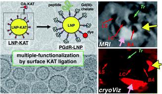
Low-density lipoprotein (LDL)-mimetic lipid nanoparticles (LNPs), decorated with MRI contrast agents and fluorescent dyes, were prepared by the covalent attachment of apolipoprotein-mimetic peptide (P), Gd(III)-chelate (Gd), and sulforhodamine B (R) moieties on the LNP surface. The functionalized LNPs were prepared using the amide-forming potassium acyltrifluoroborate (KAT) ligation reaction. The KAT groups on the surface of LNPs were allowed to react with the corresponding hydroxylamine (HA) derivatives of P and Gd to provide bi-functionalized LNPs (PGd-LNP). The reaction proceeded with excellent yields, as observed by ICP-MS (for B and Gd amounts) and MALDI-TOF-MS data, and did not alter the morphology of the LNPs (mean diameter: ca. 50 nm), as shown by DLS and cryoTEM analyses. With the help of the efficient KAT ligation, a high payload of Gd(III)-chelate on the PGd-LNP surface (ca. 2800 Gd atoms per LNP) was successfully achieved and provided a high r1 relaxivity (r1 = 22.0 s−1 mM−1 at 1.4 T/60 MHz and 25 °C; r1 = 8.2 s−1 mM−1 at 9.4 T/400 MHz and 37 °C). This bi-functionalized PGd-LNP was administered to three atherosclerotic apoE−/− mice to reveal the clear enhancement of atherosclerotic plaques in the brachiocephalic artery (BA) by MRI, in good agreement with the high accumulation of Gd in the aortic arch as shown by ICP-MS. The parallel in vivo MRI and ex vivo studies of whole mouse cryo-imaging were performed using triply functionalized LNPs with P, Gd, and R (PGdR-LNP). The clear presence of atherosclerotic plaques in BA was observed by ex vivo bright field cryo-imaging, and they were also observed by high emission fluorescent imaging. These directly corresponded to the enhanced tissue in the in vivo MRI of the identical mouse.
中文翻译:

通过表面KAT结扎制备的LDL模拟脂质纳米颗粒用于动脉粥样硬化的体内MRI
通过载脂蛋白模拟肽(P),Gd(III)-螯合物(Gd)和Gd(III)的共价连接制备了以MRI造影剂和荧光染料修饰的低密度脂蛋白(LDL)模拟脂质纳米颗粒(LNP)。LNP表面的磺基罗丹明B(R)部分。使用形成酰胺的酰基三氟硼酸钾(KAT)连接反应制备功能化的LNP。使LNP表面上的KAT基团与P和Gd的相应羟胺(HA)衍生物反应以提供双功能LNP(PGd-LNP)。如ICP-MS(对于B和Gd量)和MALDI-TOF-MS数据所观察到的,反应以优异的收率进行,并且没有改变LNP的形态(平均直径:约50 nm),如DLS和cryoTEM分析。借助有效的KAT连接,成功地在PGd-LNP表面获得了高有效载荷的Gd(III)-螯合物(每个LNP约2800 Gd原子),并提供了高r 1弛豫度(r 1 = 22.0 s -1 mM -1在1.4 T / 60 MHz和25°C时; r 1 = 8.2 s -1 mM -1在9.4 T / 400 MHz和37°C下)。如图所示,将这种双功能化的PGd-LNP给药于三只动脉粥样硬化apoE -/-小鼠,以揭示头颅动脉(BA)中动脉粥样硬化斑块的明显增强,这与Gd在主动脉弓中的高积累非常吻合通过ICP-MS 使用具有P,Gd和R的三重功能化LNP(PGdR-LNP),对整个小鼠冷冻成像进行了并行的体内MRI和离体研究。离体观察到BA中动脉粥样硬化斑块明显存在明场低温成像,也可以通过高发射荧光成像观察到。这些直接对应于同一只小鼠的体内MRI中增强的组织。
更新日期:2020-10-19
中文翻译:
通过表面KAT结扎制备的LDL模拟脂质纳米颗粒用于动脉粥样硬化的体内MRI
通过载脂蛋白模拟肽(P),Gd(III)-螯合物(Gd)和Gd(III)的共价连接制备了以MRI造影剂和荧光染料修饰的低密度脂蛋白(LDL)模拟脂质纳米颗粒(LNP)。LNP表面的磺基罗丹明B(R)部分。使用形成酰胺的酰基三氟硼酸钾(KAT)连接反应制备功能化的LNP。使LNP表面上的KAT基团与P和Gd的相应羟胺(HA)衍生物反应以提供双功能LNP(PGd-LNP)。如ICP-MS(对于B和Gd量)和MALDI-TOF-MS数据所观察到的,反应以优异的收率进行,并且没有改变LNP的形态(平均直径:约50 nm),如DLS和cryoTEM分析。借助有效的KAT连接,成功地在PGd-LNP表面获得了高有效载荷的Gd(III)-螯合物(每个LNP约2800 Gd原子),并提供了高r 1弛豫度(r 1 = 22.0 s -1 mM -1在1.4 T / 60 MHz和25°C时; r 1 = 8.2 s -1 mM -1在9.4 T / 400 MHz和37°C下)。如图所示,将这种双功能化的PGd-LNP给药于三只动脉粥样硬化apoE -/-小鼠,以揭示头颅动脉(BA)中动脉粥样硬化斑块的明显增强,这与Gd在主动脉弓中的高积累非常吻合通过ICP-MS 使用具有P,Gd和R的三重功能化LNP(PGdR-LNP),对整个小鼠冷冻成像进行了并行的体内MRI和离体研究。离体观察到BA中动脉粥样硬化斑块明显存在明场低温成像,也可以通过高发射荧光成像观察到。这些直接对应于同一只小鼠的体内MRI中增强的组织。


 京公网安备 11010802027423号