当前位置:
X-MOL 学术
›
Hum. Brain Mapp.
›
论文详情
Our official English website, www.x-mol.net, welcomes your feedback! (Note: you will need to create a separate account there.)
Computing and visualising intra‐voxel orientation‐specific relaxation–diffusion features in the human brain
Human Brain Mapping ( IF 4.8 ) Pub Date : 2020-10-06 , DOI: 10.1002/hbm.25224 João P de Almeida Martins 1, 2 , Chantal M W Tax 3, 4 , Alexis Reymbaut 1, 2 , Filip Szczepankiewicz 5, 6, 7 , Maxime Chamberland 3 , Derek K Jones 3, 8 , Daniel Topgaard 1, 2
Human Brain Mapping ( IF 4.8 ) Pub Date : 2020-10-06 , DOI: 10.1002/hbm.25224 João P de Almeida Martins 1, 2 , Chantal M W Tax 3, 4 , Alexis Reymbaut 1, 2 , Filip Szczepankiewicz 5, 6, 7 , Maxime Chamberland 3 , Derek K Jones 3, 8 , Daniel Topgaard 1, 2
Affiliation
|
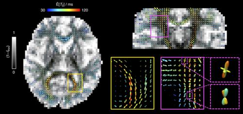
Diffusion MRI techniques are used widely to study the characteristics of the human brain connectome in vivo. However, to resolve and characterise white matter (WM) fibres in heterogeneous MRI voxels remains a challenging problem typically approached with signal models that rely on prior information and constraints. We have recently introduced a 5D relaxation–diffusion correlation framework wherein multidimensional diffusion encoding strategies are used to acquire data at multiple echo‐times to increase the amount of information encoded into the signal and ease the constraints needed for signal inversion. Nonparametric Monte Carlo inversion of the resulting datasets yields 5D relaxation–diffusion distributions where contributions from different sub‐voxel tissue environments are separated with minimal assumptions on their microscopic properties. Here, we build on the 5D correlation approach to derive fibre‐specific metrics that can be mapped throughout the imaged brain volume. Distribution components ascribed to fibrous tissues are resolved, and subsequently mapped to a dense mesh of overlapping orientation bins to define a smooth orientation distribution function (ODF). Moreover, relaxation and diffusion measures are correlated to each independent ODF coordinate, thereby allowing the estimation of orientation‐specific relaxation rates and diffusivities. The proposed method is tested on a healthy volunteer, where the estimated ODFs were observed to capture major WM tracts, resolve fibre crossings, and, more importantly, inform on the relaxation and diffusion features along with distinct fibre bundles. If combined with fibre‐tracking algorithms, the methodology presented in this work has potential for increasing the depth of characterisation of microstructural properties along individual WM pathways.
中文翻译:

计算和可视化人脑中体素内特定方向的松弛扩散特征
扩散 MRI 技术被广泛用于研究体内人脑连接组的特征。然而,解决和表征异构 MRI 体素中的白质 (WM) 纤维仍然是一个具有挑战性的问题,通常使用依赖于先验信息和约束的信号模型来解决。我们最近引入了一个 5D 弛豫-扩散相关框架,其中使用多维扩散编码策略来获取多个回波时间的数据,以增加编码到信号中的信息量并缓解信号反演所需的约束。所得数据集的非参数蒙特卡罗反演产生 5D 弛豫-扩散分布,其中来自不同亚体素组织环境的贡献被分离,对其微观特性的假设最小。在这里,我们建立在 5D 相关方法的基础上,以得出可以映射到整个成像脑容量的纤维特定指标。归因于纤维组织的分布分量被解析,并随后映射到重叠方向箱的密集网格,以定义平滑的方向分布函数 (ODF)。此外,松弛和扩散测量与每个独立的 ODF 坐标相关,从而允许估计特定方向的松弛率和扩散率。所提出的方法在一名健康志愿者身上进行了测试,其中观察到估计的 ODF 以捕获主要的 WM 束,解决纤维交叉问题,更重要的是,告知松弛和扩散特征以及不同的纤维束。如果结合光纤跟踪算法,
更新日期:2020-10-06
中文翻译:
计算和可视化人脑中体素内特定方向的松弛扩散特征
扩散 MRI 技术被广泛用于研究体内人脑连接组的特征。然而,解决和表征异构 MRI 体素中的白质 (WM) 纤维仍然是一个具有挑战性的问题,通常使用依赖于先验信息和约束的信号模型来解决。我们最近引入了一个 5D 弛豫-扩散相关框架,其中使用多维扩散编码策略来获取多个回波时间的数据,以增加编码到信号中的信息量并缓解信号反演所需的约束。所得数据集的非参数蒙特卡罗反演产生 5D 弛豫-扩散分布,其中来自不同亚体素组织环境的贡献被分离,对其微观特性的假设最小。在这里,我们建立在 5D 相关方法的基础上,以得出可以映射到整个成像脑容量的纤维特定指标。归因于纤维组织的分布分量被解析,并随后映射到重叠方向箱的密集网格,以定义平滑的方向分布函数 (ODF)。此外,松弛和扩散测量与每个独立的 ODF 坐标相关,从而允许估计特定方向的松弛率和扩散率。所提出的方法在一名健康志愿者身上进行了测试,其中观察到估计的 ODF 以捕获主要的 WM 束,解决纤维交叉问题,更重要的是,告知松弛和扩散特征以及不同的纤维束。如果结合光纤跟踪算法,


 京公网安备 11010802027423号