Journal of Neuroradiology ( IF 3.5 ) Pub Date : 2020-09-25 , DOI: 10.1016/j.neurad.2020.09.004 Maxime Ablefoni 1 , Sebastian Ullrich 1 , Alexey Surov 2 , Karl-Titus Hoffmann 3 , Hans-Jonas Meyer 1
|
Background and purpose
Diffusion-weighted imaging (DWI) is a cornerstone in diagnostic of ischemic stroke. The aim of this study was to investigate the usefulness of high-b-value computed DWI (c-DWI) in comparison to standard DWI in patients with acute brainstem infarction.
Materials and methods
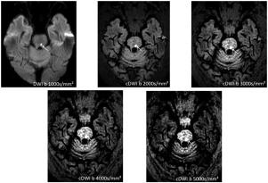
56 patients with acute brainstem infarction were retrospectively analysed by two readers. DWI was obtained with the b-values 0, 500 and 1000 s/mm² on either a 1.5 or 3 T magnetic resonance imaging (MRI) scanner. c-DWI was calculated with a monoexponential model with high b-values 2000, 3000, 4000 and 5000 s/mm². All c-DWI series with high-b-values were compared to the standard DWI sequence at b-value of 1000 s/mm² in terms of image artifacts, lesion extent and contrast.
Results
There was no statistically significant difference between 1.5 and 3 T MRI regarding the measured ischemic lesion size. There were no statistically significant differences between the ischemic lesion sizes on DWI at b-values of 1000 s/mm² and on c-DWI at higher b-values. Overall, the contrast between the lesion and the surrounding normal areas improved with increasing b-value on the isotropic DWIs: maximum at b = 5000, followed by that at b 2000 and b 1000 s/mm², in order. The best relation between artifacts and lesion contrast was identified for b 2000 s/mm².
Conclusion
High b-value DWI derived from c-DWI has a higher visibility for ischemic brainstem lesions compared to standard DWI without additional time cost. The b-2000 image is recommended to use in clinical routine, higher b-value images lead to more imaging artifacts, which might result in misdiagnosis.
中文翻译:
高b值弥散加权成像在急性脑干梗死中的诊断价值
背景和目的
弥散加权成像 (DWI) 是缺血性卒中诊断的基石。本研究的目的是调查高 b 值计算机 DWI (c-DWI) 与标准 DWI 相比在急性脑干梗死患者中的有用性。
材料和方法
两名读者对56例急性脑干梗死患者进行回顾性分析。在 1.5 或 3 T 磁共振成像 (MRI) 扫描仪上以 b 值 0、500 和 1000 s/mm² 获得 DWI。c-DWI 使用具有高 b 值 2000、3000、4000 和 5000 s/mm² 的单指数模型计算。在图像伪影、病变范围和对比度方面,将所有具有高 b 值的 c-DWI 系列与 b 值 1000 s/mm² 的标准 DWI 序列进行比较。
结果
1.5 和 3 T MRI 在测量的缺血病灶大小方面没有统计学上的显着差异。在 b 值为 1000 s/mm² 的 DWI 和较高 b 值的 c-DWI 上,缺血性病变大小之间没有统计学上的显着差异。总体而言,病变与周围正常区域之间的对比度随着各向同性 DWI 上 b 值的增加而改善:b = 5000 时最大,其次是 b 2000 和 b 1000 s/mm²,依次为。在 b 2000 s/mm² 时确定了伪影和病变对比度之间的最佳关系。
结论
与标准 DWI 相比,源自 c-DWI 的高 b 值 DWI 对缺血性脑干病变的可见度更高,无需额外的时间成本。临床常规推荐使用b-2000图像,较高的b值图像会导致更多的成像伪影,从而可能导致误诊。


 京公网安备 11010802027423号