当前位置:
X-MOL 学术
›
Eur. J. Nerosci.
›
论文详情
Our official English website, www.x-mol.net, welcomes your feedback! (Note: you will need to create a separate account there.)
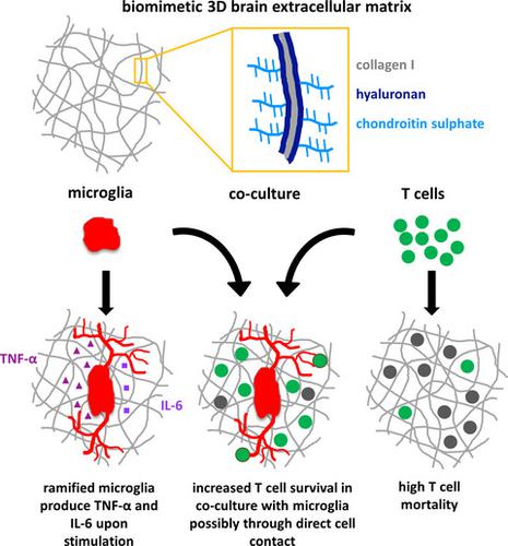
Construction of a 3D brain extracellular matrix model to study the interaction between microglia and T cells in co-culture
European Journal of Neroscience ( IF 3.4 ) Pub Date : 2020-09-20 , DOI: 10.1111/ejn.14978 Marie Frühauf 1, 2, 3 , Ulrike Zeitschel 1 , Corinna Höfling 1 , Franziska Ullm 3 , Friederike V Rabiger 2 , Gottfried Alber 2 , Tilo Pompe 3 , Uwe Müller 2 , Steffen Roßner 1
European Journal of Neroscience ( IF 3.4 ) Pub Date : 2020-09-20 , DOI: 10.1111/ejn.14978 Marie Frühauf 1, 2, 3 , Ulrike Zeitschel 1 , Corinna Höfling 1 , Franziska Ullm 3 , Friederike V Rabiger 2 , Gottfried Alber 2 , Tilo Pompe 3 , Uwe Müller 2 , Steffen Roßner 1
Affiliation
|
Neurodegenerative disorders are characterised by the activation of brain-resident microglia cells and by the infiltration of peripheral T cells. However, their interplay in disease has not been clarified yet. It is difficult to investigate complex cellular dynamics in living animals, and simple two-dimensional (2D) cell culture models do not resemble the soft 3D structure of brain tissue. Therefore, we developed a biomimetic 3D in vitro culture system for co-cultivation of microglia and T cells. As the activation and/or migration of immune cells in the brain might be affected by components of the extracellular matrix, defined 3D fibrillar collagen I-based matrices were constructed and modified with hyaluronan and/or chondroitin sulphate, resembling aspects of brain extracellular matrix. Murine microglia and spleen-derived T cells were cultured alone or in co-culture on the constructed matrices. Microglia exhibited in vivo-like morphology and T cells showed enhanced survival when co-cultured with microglia or to a minor degree in the presence of glia-conditioned medium. The open and porous fibrillar structure of the matrix allowed for cell invasion and direct cell-cell interaction, with stronger invasion of T cells. Both cell types showed no dependence on the matrix modifications. Microglia could be activated on the matrices by lipopolysaccharide resulting in interleukin-6 and tumour necrosis factor-α secretion. The findings herein indicate that biomimetic 3D matrices allow for co-cultivation and activation of primary microglia and T cells and provide useful tools to study their interaction in vitro.
中文翻译:

构建3D脑细胞外基质模型研究共培养中小胶质细胞与T细胞的相互作用
神经退行性疾病的特征是脑内小胶质细胞的激活和外周 T 细胞的浸润。然而,它们在疾病中的相互作用尚未阐明。很难研究活体动物的复杂细胞动力学,而且简单的二维 (2D) 细胞培养模型与脑组织的软 3D 结构并不相似。因此,我们开发了一种仿生 3D 体外培养系统,用于小胶质细胞和 T 细胞的共培养。由于大脑中免疫细胞的激活和/或迁移可能受到细胞外基质成分的影响,因此构建了基于 3D 纤维状胶原 I 的基质,并用透明质酸和/或硫酸软骨素进行了修饰,类似于大脑细胞外基质的各个方面。鼠小胶质细胞和脾源性 T 细胞在构建的基质上单独培养或共培养。小胶质细胞在体内表现出类似的形态,T 细胞在与小胶质细胞共培养时或在胶质细胞条件培养基的存在下以较小的程度显示出更高的存活率。基质的开放和多孔的纤维状结构允许细胞侵袭和直接的细胞间相互作用,T细胞的侵袭能力更强。两种细胞类型均不依赖于基质修饰。脂多糖可以激活基质上的小胶质细胞,导致白细胞介素 6 和肿瘤坏死因子 α 的分泌。本文的研究结果表明,仿生 3D 矩阵允许原代小胶质细胞和 T 细胞的共培养和激活,并提供有用的工具来研究它们的体外相互作用。
更新日期:2020-09-20
中文翻译:
构建3D脑细胞外基质模型研究共培养中小胶质细胞与T细胞的相互作用
神经退行性疾病的特征是脑内小胶质细胞的激活和外周 T 细胞的浸润。然而,它们在疾病中的相互作用尚未阐明。很难研究活体动物的复杂细胞动力学,而且简单的二维 (2D) 细胞培养模型与脑组织的软 3D 结构并不相似。因此,我们开发了一种仿生 3D 体外培养系统,用于小胶质细胞和 T 细胞的共培养。由于大脑中免疫细胞的激活和/或迁移可能受到细胞外基质成分的影响,因此构建了基于 3D 纤维状胶原 I 的基质,并用透明质酸和/或硫酸软骨素进行了修饰,类似于大脑细胞外基质的各个方面。鼠小胶质细胞和脾源性 T 细胞在构建的基质上单独培养或共培养。小胶质细胞在体内表现出类似的形态,T 细胞在与小胶质细胞共培养时或在胶质细胞条件培养基的存在下以较小的程度显示出更高的存活率。基质的开放和多孔的纤维状结构允许细胞侵袭和直接的细胞间相互作用,T细胞的侵袭能力更强。两种细胞类型均不依赖于基质修饰。脂多糖可以激活基质上的小胶质细胞,导致白细胞介素 6 和肿瘤坏死因子 α 的分泌。本文的研究结果表明,仿生 3D 矩阵允许原代小胶质细胞和 T 细胞的共培养和激活,并提供有用的工具来研究它们的体外相互作用。


 京公网安备 11010802027423号