当前位置:
X-MOL 学术
›
J. Leukoc. Biol.
›
论文详情
Our official English website, www.x-mol.net, welcomes your feedback! (Note: you will need to create a separate account there.)
Sterile inflammation drives multiple programmed cell death pathways in the gut.
Journal of Leukocyte Biology ( IF 5.5 ) Pub Date : 2020-09-18 , DOI: 10.1002/jlb.3ma0820-660r Carolina N Ruera 1 , Emanuel Miculán 1 , Federico Pérez 1 , Gerónimo Ducca 1 , Paula Carasi 1 , Fernando G Chirdo 1
Journal of Leukocyte Biology ( IF 5.5 ) Pub Date : 2020-09-18 , DOI: 10.1002/jlb.3ma0820-660r Carolina N Ruera 1 , Emanuel Miculán 1 , Federico Pérez 1 , Gerónimo Ducca 1 , Paula Carasi 1 , Fernando G Chirdo 1
Affiliation

|
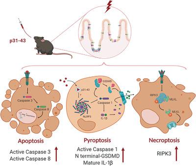
Intestinal epithelial cells have a rapid turnover, being rapidly renewed by newly differentiated enterocytes, balanced by massive and constant removal of damaged cells by programmed cell death (PCD). The main forms of PCD are apoptosis, pyroptosis, and necroptosis, with apoptosis being a noninflammatory process, whereas the others drive innate immune responses. Although apoptosis is thought to be the principal means of cell death in the healthy intestine, which mechanisms are responsible for PCD during inflammation are not fully understood. To address this question, we used an in vivo model of enteropathy in wild‐type mice induced by a single intragastric administration of the p31‐43 gliadin peptide, which is known to elicit transient MyD88, NLRP3, and caspase‐1‐dependent mucosal damage and inflammation in the small intestine. Here, we found increased numbers of TUNEL+ cells in the mucosa as early as 2 h after p31‐43 administration. Western blot and immunofluorescence analysis showed the presence of caspase‐3‐mediated apoptosis in the epithelium and lamina propria. In addition, the presence of mature forms of caspase‐1, IL‐1β, and gasdermin D showed activation of pyroptosis and inhibition of caspase‐1 led to decreased enterocyte death in p31‐43‐treated mice. There was also up‐regulation of RIPK3 in crypt epithelium, suggesting that necroptosis was also occurring. Taken together, these results indicate that the inflammatory response induced by p31‐43 can drive multiple PCD pathways in the small intestine.
中文翻译:

无菌炎症驱动肠道中多个程序性细胞死亡途径。
肠上皮细胞具有快速更新,被新分化的肠上皮细胞快速更新,与通过程序性细胞死亡(PCD)不断大量清除受损细胞而达到平衡的功能。PCD的主要形式是凋亡,凋亡和坏死性坏死,凋亡是非炎性过程,而其他形式则驱动先天性免疫应答。尽管凋亡被认为是健康肠道中细胞死亡的主要手段,但炎症过程中导致PCD的机制尚不完全清楚。为了解决这个问题,我们使用了一次胃内施用p31-43醇溶蛋白肽诱导的野生型小鼠肠病的体内模型,已知该蛋白会引起短暂的MyD88,NLRP3和caspase-1依赖性粘膜损伤和小肠发炎。这里,施用p31-43后2小时内粘膜中的+细胞。Western印迹和免疫荧光分析显示上皮和固有层中存在caspase-3介导的凋亡。此外,成熟形式的caspase-1,IL-1β和gasdermin D的存在表明激活了细胞凋亡,而caspase-1的抑制导致p31-43处理的小鼠肠细胞死亡减少。隐窝上皮细胞中的RIPK3也上调,提示坏死也正在发生。综上所述,这些结果表明,p31-43诱导的炎症反应可以驱动小肠中的多个PCD途径。
更新日期:2020-09-18
中文翻译:

无菌炎症驱动肠道中多个程序性细胞死亡途径。
肠上皮细胞具有快速更新,被新分化的肠上皮细胞快速更新,与通过程序性细胞死亡(PCD)不断大量清除受损细胞而达到平衡的功能。PCD的主要形式是凋亡,凋亡和坏死性坏死,凋亡是非炎性过程,而其他形式则驱动先天性免疫应答。尽管凋亡被认为是健康肠道中细胞死亡的主要手段,但炎症过程中导致PCD的机制尚不完全清楚。为了解决这个问题,我们使用了一次胃内施用p31-43醇溶蛋白肽诱导的野生型小鼠肠病的体内模型,已知该蛋白会引起短暂的MyD88,NLRP3和caspase-1依赖性粘膜损伤和小肠发炎。这里,施用p31-43后2小时内粘膜中的+细胞。Western印迹和免疫荧光分析显示上皮和固有层中存在caspase-3介导的凋亡。此外,成熟形式的caspase-1,IL-1β和gasdermin D的存在表明激活了细胞凋亡,而caspase-1的抑制导致p31-43处理的小鼠肠细胞死亡减少。隐窝上皮细胞中的RIPK3也上调,提示坏死也正在发生。综上所述,这些结果表明,p31-43诱导的炎症反应可以驱动小肠中的多个PCD途径。


 京公网安备 11010802027423号