当前位置:
X-MOL 学术
›
Anal. Chem.
›
论文详情
Our official English website, www.x-mol.net, welcomes your feedback! (Note: you will need to create a separate account there.)
Raman spectroscopy with 2D perturbation correlation moving windows for the characterisation of heparin-amyloid interactions.
Analytical Chemistry ( IF 7.4 ) Pub Date : 2020-09-16 , DOI: 10.1021/acs.analchem.0c02390 David J Townsend 1 , David A Middleton 1 , Lorna Ashton 1
Analytical Chemistry ( IF 7.4 ) Pub Date : 2020-09-16 , DOI: 10.1021/acs.analchem.0c02390 David J Townsend 1 , David A Middleton 1 , Lorna Ashton 1
Affiliation

|
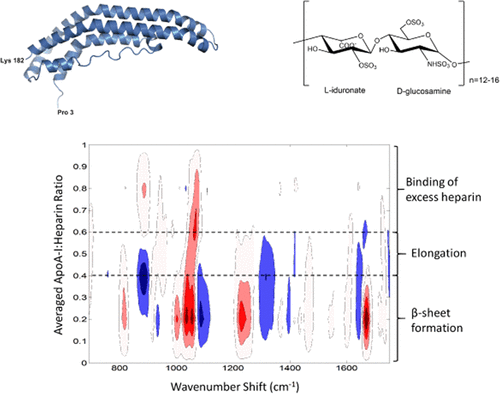
It has been shown extensively that glycosaminoglycan (GAG)–protein interactions can induce, accelerate, and impede the clearance of amyloid fibrils associated with systemic and localized amyloidosis. Obtaining molecular details of these interactions is fundamental to our understanding of amyloid disease. Consequently, there is a need for analytical approaches that can identify protein conformational transitions and simultaneously characterize heparin interactions. By combining Raman spectroscopy with two-dimensional (2D) perturbation correlation moving window (2DPCMW) analysis, we have successfully identified changes in protein secondary structure during pH- and heparin-induced fibril formation of apolipoprotein A-I (apoA-I) associated with atherosclerosis. Furthermore, from the 2DPCMW, we have identified peak shifts and intensity variations in Raman peaks arising from different heparan sulfate moieties, indicating that protein–heparin interactions vary at different heparin concentrations. Raman spectroscopy thus reveals new mechanistic insights into the role of GAGs during amyloid fibril formation.
中文翻译:

拉曼光谱具有二维微扰相关移动窗口,用于表征肝素-淀粉样蛋白相互作用。
大量研究表明,糖胺聚糖(GAG)-蛋白质相互作用可以诱导,加速和阻止与全身性和局部性淀粉样变性有关的淀粉样原纤维的清除。获得这些相互作用的分子细节是我们对淀粉样蛋白疾病理解的基础。因此,需要能够识别蛋白质构象转变并同时表征肝素相互作用的分析方法。通过将拉曼光谱与二维(2D)扰动相关移动窗口(2DPCMW)分析相结合,我们已成功识别了在pH和肝素诱导的与动脉粥样硬化相关的载脂蛋白AI(apoA-I)的原纤维形成过程中蛋白质二级结构的变化。此外,从2DPCMW 我们已经确定了硫酸乙酰肝素不同部分引起的拉曼峰的峰移动和强度变化,表明蛋白质-肝素相互作用在不同肝素浓度下会发生变化。拉曼光谱因此揭示了在淀粉样蛋白原纤维形成过程中GAG的作用的新机制。
更新日期:2020-10-21
中文翻译:

拉曼光谱具有二维微扰相关移动窗口,用于表征肝素-淀粉样蛋白相互作用。
大量研究表明,糖胺聚糖(GAG)-蛋白质相互作用可以诱导,加速和阻止与全身性和局部性淀粉样变性有关的淀粉样原纤维的清除。获得这些相互作用的分子细节是我们对淀粉样蛋白疾病理解的基础。因此,需要能够识别蛋白质构象转变并同时表征肝素相互作用的分析方法。通过将拉曼光谱与二维(2D)扰动相关移动窗口(2DPCMW)分析相结合,我们已成功识别了在pH和肝素诱导的与动脉粥样硬化相关的载脂蛋白AI(apoA-I)的原纤维形成过程中蛋白质二级结构的变化。此外,从2DPCMW 我们已经确定了硫酸乙酰肝素不同部分引起的拉曼峰的峰移动和强度变化,表明蛋白质-肝素相互作用在不同肝素浓度下会发生变化。拉曼光谱因此揭示了在淀粉样蛋白原纤维形成过程中GAG的作用的新机制。


 京公网安备 11010802027423号