当前位置:
X-MOL 学术
›
J. Biophotonics
›
论文详情
Our official English website, www.x-mol.net, welcomes your feedback! (Note: you will need to create a separate account there.)
Detecting mouse squamous cell carcinoma from submicron full-field optical coherence tomography images by deep learning.
Journal of Biophotonics ( IF 2.8 ) Pub Date : 2020-09-05 , DOI: 10.1002/jbio.202000271 Chi-Jui Ho,Manuel Calderon-Delgado,Chin-Cheng Chan,Ming-Yi Lin,Jeng-Wei Tjiu,Sheng-Lung Huang,Homer H Chen
Journal of Biophotonics ( IF 2.8 ) Pub Date : 2020-09-05 , DOI: 10.1002/jbio.202000271 Chi-Jui Ho,Manuel Calderon-Delgado,Chin-Cheng Chan,Ming-Yi Lin,Jeng-Wei Tjiu,Sheng-Lung Huang,Homer H Chen
|
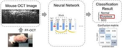
The standard medical practice for cancer diagnosis requires histopathology, which is an invasive and time‐consuming procedure. Optical coherence tomography (OCT) is an alternative that is relatively fast, noninvasive, and able to capture three‐dimensional structures of epithelial tissue. Unlike most previous OCT systems, which cannot capture crucial cellular‐level information for squamous cell carcinoma (SCC) diagnosis, the full‐field OCT (FF‐OCT) technology used in this paper is able to produce images at sub‐micron resolution and thereby facilitates the development of a deep learning algorithm for SCC detection. Experimental results show that the SCC detection algorithm can achieve a classification accuracy of 80% for mouse skin. Using the sub‐micron FF‐OCT imaging system, the proposed SCC detection algorithm has the potential for in‐vivo applications.
中文翻译:

通过深度学习从亚微米全场光学相干断层扫描图像中检测小鼠鳞状细胞癌。
用于癌症诊断的标准医学实践需要组织病理学,这是一种侵入性且耗时的过程。光学相干断层扫描(OCT)是一种相对快速,无创且能够捕获上皮组织的三维结构的替代方法。与大多数以前的OCT系统无法捕获鳞状细胞癌(SCC)诊断的关键细胞水平信息不同,本文使用的全场OCT(FF-OCT)技术能够产生亚微米分辨率的图像,从而有助于开发用于SCC检测的深度学习算法。实验结果表明,SCC检测算法可以对小鼠皮肤实现80%的分类精度。使用亚微米FF-OCT成像系统,
更新日期:2020-09-05
中文翻译:
通过深度学习从亚微米全场光学相干断层扫描图像中检测小鼠鳞状细胞癌。
用于癌症诊断的标准医学实践需要组织病理学,这是一种侵入性且耗时的过程。光学相干断层扫描(OCT)是一种相对快速,无创且能够捕获上皮组织的三维结构的替代方法。与大多数以前的OCT系统无法捕获鳞状细胞癌(SCC)诊断的关键细胞水平信息不同,本文使用的全场OCT(FF-OCT)技术能够产生亚微米分辨率的图像,从而有助于开发用于SCC检测的深度学习算法。实验结果表明,SCC检测算法可以对小鼠皮肤实现80%的分类精度。使用亚微米FF-OCT成像系统,


 京公网安备 11010802027423号