当前位置:
X-MOL 学术
›
J. Neurochem.
›
论文详情
Our official English website, www.x-mol.net, welcomes your feedback! (Note: you will need to create a separate account there.)
Characterization and heme oxygenase-1 content of extracellular vesicles in human biofluids
Journal of Neurochemistry ( IF 4.7 ) Pub Date : 2020-09-03 , DOI: 10.1111/jnc.15167 Marisa Cressatti 1, 2 , Julia M Galindez 1, 2 , Lamin Juwara 1, 3 , Natalie Orlovetskie 1, 2 , Ana M Velly 1, 4, 5 , Shaun Eintracht 6 , Adrienne Liberman 1 , Mervyn Gornitsky 1, 4, 5 , Hyman M Schipper 1, 2
Journal of Neurochemistry ( IF 4.7 ) Pub Date : 2020-09-03 , DOI: 10.1111/jnc.15167 Marisa Cressatti 1, 2 , Julia M Galindez 1, 2 , Lamin Juwara 1, 3 , Natalie Orlovetskie 1, 2 , Ana M Velly 1, 4, 5 , Shaun Eintracht 6 , Adrienne Liberman 1 , Mervyn Gornitsky 1, 4, 5 , Hyman M Schipper 1, 2
Affiliation
|
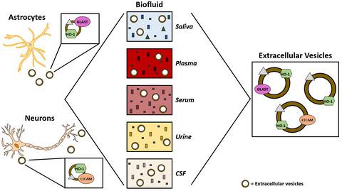
Heme oxygenase-1 (HO-1), a highly inducible stress protein that degrades heme to biliverdin, carbon monoxide, and free ferrous iron, is increased in blood and other biofluids of subjects with various systemic and neurological disorders. HO-1 does not contain an N-terminal signal peptide and the mechanism responsible for its secretion remains unknown. Extracellular vesicles (EVs) are membrane-bound inclusions that transport microRNAs, messenger RNAs, lipids, and proteins among diverse cellular and extracellular compartments. The objective of the current study was to determine whether EVs in human biofluids contain HO-1, and whether the latter may be transported in EVs from brain to periphery. Total, L1 cell adhesion molecule protein (L1CAM)-enriched (neuron-derived), and glutamate aspartate transporter 1 (GLAST)-enriched (astrocyte-derived) EVs were purified from five different human biofluids (saliva [n = 40], plasma [n = 14], serum [n = 10], urine [n = 10], and cerebrospinal fluid [n = 11]) using polymer precipitation and immuno-affinity-based capture methods. L1CAM-enriched, GLAST-enriched, and L1CAM/GLAST-depleted (LGD) EV, along with EV-depleted (EVD), fractions were validated by nanoparticle tracking analysis, enzyme-linked immunosorbent assay (ELISA), and western blot. HO-1 was assayed in all fractions using ELISA and western blot. The majority of HO-1 protein was localized to LGD, L1CAM-enriched, and GLAST-enriched EVs of all human biofluids surveyed after adjusting for age and sex, with little HO-1 protein detected in EVD fractions. HO-1 protein in human biofluids is predominantly localized to EV compartments. A substantial proportion of EV HO-1 in peripheral human biofluids is derived from the central nervous system and may contribute to the systemic manifestations of various neurological conditions.
中文翻译:

人体生物体液中细胞外囊泡的表征和血红素加氧酶-1的含量
血红素加氧酶-1 (HO-1) 是一种高度诱导性应激蛋白,可将血红素降解为胆绿素、一氧化碳和游离亚铁,在患有各种全身性和神经系统疾病的受试者的血液和其他生物体液中增加。HO-1 不含 N 端信号肽,其分泌机制尚不清楚。细胞外囊泡 (EV) 是膜结合的内含物,可在不同的细胞和细胞外隔室之间运输 microRNA、信使 RNA、脂质和蛋白质。当前研究的目的是确定人类生物体液中的 EV 是否含有 HO-1,以及后者是否可以在 EV 中从大脑运输到外周。总计,富含 L1 细胞粘附分子蛋白 (L1CAM)(神经元衍生),n = 40]、血浆 [ n = 14]、血清 [ n = 10]、尿液 [ n = 10] 和脑脊液 [ n = 11]) 使用聚合物沉淀和基于免疫亲和的捕获方法。L1CAM 富集、GLAST 富集和 L1CAM/GLAST 耗尽 (LGD) EV,连同 EV 耗尽 (EVD),通过纳米粒子追踪分析、酶联免疫吸附测定 (ELISA) 和蛋白质印迹验证了级分。使用 ELISA 和蛋白质印迹法测定所有级分中的 HO-1。大多数 HO-1 蛋白定位于 LGD、富含 L1CAM 和富含 GLAST 的所有人类生物体液的 EV,在调整年龄和性别后进行调查,在 EVD 级分中检测到的 HO-1 蛋白很少。人类生物体液中的 HO-1 蛋白主要定位于 EV 隔室。外周人体生物体液中很大一部分 EV HO-1 来自中枢神经系统,可能导致各种神经系统疾病的全身表现。
更新日期:2020-09-03
中文翻译:
人体生物体液中细胞外囊泡的表征和血红素加氧酶-1的含量
血红素加氧酶-1 (HO-1) 是一种高度诱导性应激蛋白,可将血红素降解为胆绿素、一氧化碳和游离亚铁,在患有各种全身性和神经系统疾病的受试者的血液和其他生物体液中增加。HO-1 不含 N 端信号肽,其分泌机制尚不清楚。细胞外囊泡 (EV) 是膜结合的内含物,可在不同的细胞和细胞外隔室之间运输 microRNA、信使 RNA、脂质和蛋白质。当前研究的目的是确定人类生物体液中的 EV 是否含有 HO-1,以及后者是否可以在 EV 中从大脑运输到外周。总计,富含 L1 细胞粘附分子蛋白 (L1CAM)(神经元衍生),n = 40]、血浆 [ n = 14]、血清 [ n = 10]、尿液 [ n = 10] 和脑脊液 [ n = 11]) 使用聚合物沉淀和基于免疫亲和的捕获方法。L1CAM 富集、GLAST 富集和 L1CAM/GLAST 耗尽 (LGD) EV,连同 EV 耗尽 (EVD),通过纳米粒子追踪分析、酶联免疫吸附测定 (ELISA) 和蛋白质印迹验证了级分。使用 ELISA 和蛋白质印迹法测定所有级分中的 HO-1。大多数 HO-1 蛋白定位于 LGD、富含 L1CAM 和富含 GLAST 的所有人类生物体液的 EV,在调整年龄和性别后进行调查,在 EVD 级分中检测到的 HO-1 蛋白很少。人类生物体液中的 HO-1 蛋白主要定位于 EV 隔室。外周人体生物体液中很大一部分 EV HO-1 来自中枢神经系统,可能导致各种神经系统疾病的全身表现。


 京公网安备 11010802027423号