当前位置:
X-MOL 学术
›
Eur. Polym. J.
›
论文详情
Our official English website, www.x-mol.net, welcomes your feedback! (Note: you will need to create a separate account there.)
3D Scaffolding of fast photocurable polyurethane for soft tissue engineering by stereolithography: Influence of materials and geometry on growth of fibroblast cells
European Polymer Journal ( IF 6 ) Pub Date : 2020-10-01 , DOI: 10.1016/j.eurpolymj.2020.109988 Afsoon Farzan , Sedigheh Borandeh , Nazanin Zanjanizadeh Ezazi , Sami Lipponen , Hélder A. Santos , Jukka Seppälä
European Polymer Journal ( IF 6 ) Pub Date : 2020-10-01 , DOI: 10.1016/j.eurpolymj.2020.109988 Afsoon Farzan , Sedigheh Borandeh , Nazanin Zanjanizadeh Ezazi , Sami Lipponen , Hélder A. Santos , Jukka Seppälä
|
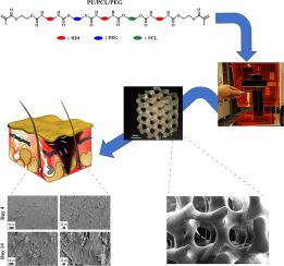
Abstract Tissue engineering can benefit from the availability of three-dimensional (3D) printing technologies that make it possible to produce scaffolds with complex geometry. Chemical, mechanical, and structural properties should be considered in scaffold design and development since these properties affect cell adhesion, proliferation, and differentiation. To this end, in this study, we developed a series of fast photocuring polyurethanes (PUs), using poly(e-caprolactone) (PCL) and/or polyethylene glycol (PEG) as microdiols, using a solvent-free method and stereolithography strategy for the fabrication of elastic 3D-printed scaffold. The effects of different diols on the hydrolytic degradation, thermal and mechanical properties, and hydrophilicity of PUs were evaluated. The results showed that PEG-containing PUs had higher degradation rates, and the tensile strength of PU/PCL/PEG was 1.4 and 2 times higher than that of PU/PEG and PU/PCL, respectively. Moreover, the effect of different diols and scaffold geometry on toxicity and cell attachment were studied in vitro. The results of MTT and AlamarBlue assays on dermal fibroblast cells showed high proliferation of printed PU/PCL/PEG scaffold with no sign of cytotoxicity. In addition, compared to cast film PUs, relatively high cell attachment was seen on the surface of printed PU/PCL/PEG even after 4 days. Therefore, 3D printed PU/PCL/PEG showed high applicability in soft tissue engineering, especially for scaffold development.
中文翻译:

立体光刻法用于软组织工程的快速光固化聚氨酯的 3D 支架:材料和几何形状对成纤维细胞生长的影响
摘要 组织工程可以受益于三维 (3D) 打印技术的可用性,该技术使得生产具有复杂几何形状的支架成为可能。在支架设计和开发中应考虑化学、机械和结构特性,因为这些特性会影响细胞粘附、增殖和分化。为此,在本研究中,我们开发了一系列快速光固化聚氨酯(PU),使用聚(ε-己内酯)(PCL)和/或聚乙二醇(PEG)作为微二醇,使用无溶剂方法和立体光刻策略用于制造弹性 3D 打印支架。评估了不同二醇对聚氨酯的水解降解、热和机械性能以及亲水性的影响。结果表明,含 PEG 的 PU 具有更高的降解率,PU/PCL/PEG 的拉伸强度分别是 PU/PEG 和 PU/PCL 的 1.4 倍和 2 倍。此外,在体外研究了不同二醇和支架几何形状对毒性和细胞附着的影响。MTT 和 AlamarBlue 对真皮成纤维细胞的测定结果显示打印的 PU/PCL/PEG 支架具有高增殖,没有细胞毒性迹象。此外,与流延膜 PU 相比,即使在 4 天后,在印刷的 PU/PCL/PEG 表面上也能看到相对较高的细胞附着。因此,3D 打印的 PU/PCL/PEG 在软组织工程中表现出很高的适用性,尤其是在支架开发方面。体外研究了不同二醇和支架几何形状对毒性和细胞附着的影响。MTT 和 AlamarBlue 对真皮成纤维细胞的测定结果显示打印的 PU/PCL/PEG 支架具有高增殖,没有细胞毒性迹象。此外,与流延膜 PU 相比,即使在 4 天后,在印刷的 PU/PCL/PEG 表面上也能看到相对较高的细胞附着。因此,3D 打印的 PU/PCL/PEG 在软组织工程中表现出很高的适用性,尤其是在支架开发方面。体外研究了不同二醇和支架几何形状对毒性和细胞附着的影响。MTT 和 AlamarBlue 对真皮成纤维细胞的测定结果显示打印的 PU/PCL/PEG 支架具有高增殖,没有细胞毒性迹象。此外,与流延膜 PU 相比,即使在 4 天后,在印刷的 PU/PCL/PEG 表面上也能看到相对较高的细胞附着。因此,3D 打印的 PU/PCL/PEG 在软组织工程中表现出很高的适用性,尤其是在支架开发方面。
更新日期:2020-10-01
中文翻译:
立体光刻法用于软组织工程的快速光固化聚氨酯的 3D 支架:材料和几何形状对成纤维细胞生长的影响
摘要 组织工程可以受益于三维 (3D) 打印技术的可用性,该技术使得生产具有复杂几何形状的支架成为可能。在支架设计和开发中应考虑化学、机械和结构特性,因为这些特性会影响细胞粘附、增殖和分化。为此,在本研究中,我们开发了一系列快速光固化聚氨酯(PU),使用聚(ε-己内酯)(PCL)和/或聚乙二醇(PEG)作为微二醇,使用无溶剂方法和立体光刻策略用于制造弹性 3D 打印支架。评估了不同二醇对聚氨酯的水解降解、热和机械性能以及亲水性的影响。结果表明,含 PEG 的 PU 具有更高的降解率,PU/PCL/PEG 的拉伸强度分别是 PU/PEG 和 PU/PCL 的 1.4 倍和 2 倍。此外,在体外研究了不同二醇和支架几何形状对毒性和细胞附着的影响。MTT 和 AlamarBlue 对真皮成纤维细胞的测定结果显示打印的 PU/PCL/PEG 支架具有高增殖,没有细胞毒性迹象。此外,与流延膜 PU 相比,即使在 4 天后,在印刷的 PU/PCL/PEG 表面上也能看到相对较高的细胞附着。因此,3D 打印的 PU/PCL/PEG 在软组织工程中表现出很高的适用性,尤其是在支架开发方面。体外研究了不同二醇和支架几何形状对毒性和细胞附着的影响。MTT 和 AlamarBlue 对真皮成纤维细胞的测定结果显示打印的 PU/PCL/PEG 支架具有高增殖,没有细胞毒性迹象。此外,与流延膜 PU 相比,即使在 4 天后,在印刷的 PU/PCL/PEG 表面上也能看到相对较高的细胞附着。因此,3D 打印的 PU/PCL/PEG 在软组织工程中表现出很高的适用性,尤其是在支架开发方面。体外研究了不同二醇和支架几何形状对毒性和细胞附着的影响。MTT 和 AlamarBlue 对真皮成纤维细胞的测定结果显示打印的 PU/PCL/PEG 支架具有高增殖,没有细胞毒性迹象。此外,与流延膜 PU 相比,即使在 4 天后,在印刷的 PU/PCL/PEG 表面上也能看到相对较高的细胞附着。因此,3D 打印的 PU/PCL/PEG 在软组织工程中表现出很高的适用性,尤其是在支架开发方面。


 京公网安备 11010802027423号