International Journal for Parasitology ( IF 4 ) Pub Date : 2020-09-01 , DOI: 10.1016/j.ijpara.2020.07.007 Ha Ngo-Thanh 1 , Tsutomu Sasaki 2 , Kazutomo Suzue 1 , Hideaki Yokoo 3 , Koji Isoda 3 , Wataru Kamitani 4 , Chikako Shimokawa 5 , Hajime Hisaeda 5 , Takashi Imai 1
|
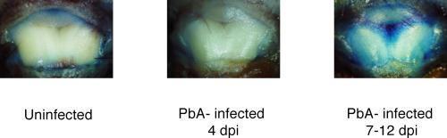
Cerebral malaria is one of the most severe pathologies of malaria; it induces neuro-cognitive sequelae and has a high mortality rate. Although many factors involved in the development of cerebral malaria have been discovered, its pathogenic mechanisms are still not completely understood. Most studies on cerebral malaria have focused on the blood–brain barrier, despite the importance of the blood–cerebrospinal fluid barrier, which protects the brain from peripheral inflammation. Consequently, the pathological role of the blood–cerebrospinal fluid barrier in cerebral malaria is currently unknown. To examine the status of the blood–cerebrospinal fluid barrier in cerebral malaria and malaria without this pathology (non-cerebral malaria), we developed a new method for evaluating the permeabilization of the blood–cerebrospinal fluid barrier during cerebral malaria in mice, using Evans blue dye and a software-assisted image analysis. Using C57BL/6J (B6) mice infected with Plasmodium berghei ANKA strain as an experimental cerebral malaria model and B6 mice infected with P. berghei NK65 strain or Plasmodium yoelii as non-cerebral malaria models, we revealed that the permeability of the blood–cerebrospinal fluid barrier increased during experimental cerebral malaria but not during non-cerebral malaria. We observed haemorrhaging in the cerebral ventricles and hemozoin-like structures in the choroid plexus, which is a key component of the blood–cerebrospinal fluid barrier, in cerebral malaria mice. Taken together, this evidence indicates that the blood–cerebrospinal fluid barrier is disrupted in experimental cerebral malaria, whereas it remains intact in non-cerebral malaria. We also found that P. berghei ANKA parasites and CD8+ T cells are involved in the blood–cerebrospinal fluid barrier disruption in experimental cerebral malaria. An understanding of the mechanisms underlying cerebral malaria might help in the development of effective strategies to prevent and manage cerebral malaria in humans.
中文翻译:
血脑脊液屏障:在由伯氏疟原虫 ANKA 引起的实验性脑疟疾期间被破坏的另一个部位。
脑型疟疾是疟疾最严重的疾病之一;它会诱发神经认知后遗症,并且死亡率很高。尽管已经发现了许多参与脑型疟疾发展的因素,但其发病机制仍未完全了解。尽管血脑脊液屏障保护大脑免受外周炎症的影响,但大多数关于脑型疟疾的研究都集中在血脑屏障上。因此,血脑脊液屏障在脑型疟疾中的病理作用目前尚不清楚。为了检查脑型疟疾和没有这种病理的疟疾(非脑型疟疾)中血脑脊液屏障的状态,我们开发了一种新方法,用于评估小鼠脑型疟疾期间血脑脊液屏障的通透性,使用伊文思蓝染料和软件辅助图像分析。使用 C57BL/6J (B6) 小鼠感染伯氏疟原虫ANKA 菌株作为实验性脑疟疾模型和感染P 的B6 小鼠。berghei NK65 菌株或约氏疟原虫作为非脑型疟疾模型,我们发现在实验性脑型疟疾期间血脑脊液屏障的通透性增加,但在非脑型疟疾期间没有增加。我们在脑疟疾小鼠中观察到脑室出血和脉络丛中的血红素样结构,脉络丛是血脑脊液屏障的关键组成部分。综上所述,这一证据表明,在实验性脑型疟疾中,血脑脊液屏障被破坏,而在非脑型疟疾中则保持完整。我们还发现P. berghei ANKA 寄生虫和 CD8 + T 细胞参与实验性脑疟疾的血脑脊液屏障破坏。了解脑型疟疾的潜在机制可能有助于制定预防和管理人类脑型疟疾的有效策略。


 京公网安备 11010802027423号