Our official English website, www.x-mol.net, welcomes your feedback! (Note: you will need to create a separate account there.)
Machine vision-driven automatic recognition of particle size and morphology in SEM images.
Nanoscale ( IF 6.7 ) Pub Date : 2020-08-27 , DOI: 10.1039/d0nr04140h Hyojin Kim 1 , Jinkyu Han 2 , T Yong-Jin Han 2
Nanoscale ( IF 6.7 ) Pub Date : 2020-08-27 , DOI: 10.1039/d0nr04140h Hyojin Kim 1 , Jinkyu Han 2 , T Yong-Jin Han 2
Affiliation
|
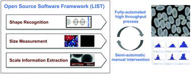
Scanning Electron Microscopy (SEM) images provide a variety of structural and morphological information of nanomaterials. In the material informatics domain, automatic recognition and quantitative analysis of SEM images in a high-throughput manner are critical, but challenges still remain due to the complexity and the diversity of image configurations in both shape and size. In this paper, we present a generally applicable approach using computer vision and machine learning techniques to quantitatively extract particle size, size distribution and morphology information in SEM images. The proposed pipeline offers automatic, high-throughput measurements even when overlapping nanoparticles, rod shapes, and core–shell nanostructures are present. We demonstrate effectiveness of the proposed approach by performing experiments on SEM images of nanoscale materials and structures with different shapes and sizes. The proposed approach shows promising results (Spearman coefficients of 0.91 and 0.99 using fully automated and semi-automated processes, respectively) when compared with manually measured sizes. The code is made available as open source software at https://github.com/LLNL/LIST.
中文翻译:

机器视觉驱动的SEM图像中颗粒大小和形态的自动识别。
扫描电子显微镜(SEM)图像提供了纳米材料的各种结构和形态信息。在材料信息学领域,以高通量方式对SEM图像进行自动识别和定量分析至关重要,但是由于形状和尺寸上图像配置的复杂性和多样性,仍然存在挑战。在本文中,我们提出一种使用计算机视觉和机器学习技术的通用方法,以定量提取SEM图像中的粒径,粒径分布和形态信息。即使存在重叠的纳米颗粒,棒状形状和核-壳纳米结构,建议的管道也可提供自动的高通量测量。我们通过对具有不同形状和大小的纳米级材料和结构的SEM图像进行实验,证明了该方法的有效性。与手动测量的尺寸相比,所提出的方法显示出令人鼓舞的结果(分别使用全自动和半自动化过程的Spearman系数分别为0.91和0.99)。该代码可作为开源软件在https://github.com/LLNL/LIST上获得。
更新日期:2020-10-02
中文翻译:
机器视觉驱动的SEM图像中颗粒大小和形态的自动识别。
扫描电子显微镜(SEM)图像提供了纳米材料的各种结构和形态信息。在材料信息学领域,以高通量方式对SEM图像进行自动识别和定量分析至关重要,但是由于形状和尺寸上图像配置的复杂性和多样性,仍然存在挑战。在本文中,我们提出一种使用计算机视觉和机器学习技术的通用方法,以定量提取SEM图像中的粒径,粒径分布和形态信息。即使存在重叠的纳米颗粒,棒状形状和核-壳纳米结构,建议的管道也可提供自动的高通量测量。我们通过对具有不同形状和大小的纳米级材料和结构的SEM图像进行实验,证明了该方法的有效性。与手动测量的尺寸相比,所提出的方法显示出令人鼓舞的结果(分别使用全自动和半自动化过程的Spearman系数分别为0.91和0.99)。该代码可作为开源软件在https://github.com/LLNL/LIST上获得。


 京公网安备 11010802027423号