当前位置:
X-MOL 学术
›
J. Biophotonics
›
论文详情
Our official English website, www.x-mol.net, welcomes your feedback! (Note: you will need to create a separate account there.)
In situ measurement of the isoplanatic patch for imaging through intact bone.
Journal of Biophotonics ( IF 2.8 ) Pub Date : 2020-08-25 , DOI: 10.1002/jbio.202000160 Kayvan Forouhesh Tehrani 1 , Nektarios Koukourakis 2, 3 , Jürgen Czarske 2, 3, 4 , Luke J Mortensen 1, 5
Journal of Biophotonics ( IF 2.8 ) Pub Date : 2020-08-25 , DOI: 10.1002/jbio.202000160 Kayvan Forouhesh Tehrani 1 , Nektarios Koukourakis 2, 3 , Jürgen Czarske 2, 3, 4 , Luke J Mortensen 1, 5
Affiliation

|
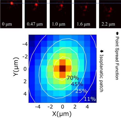
Wavefront‐shaping (WS) enables imaging through scattering tissues like bone, which is important for neuroscience and bone‐regeneration research. WS corrects for the optical aberrations at a given depth and field‐of‐view (FOV) within the sample; the extent of the validity of which is limited to a region known as the isoplanatic patch (IP). Knowing this parameter helps to estimate the number of corrections needed for WS imaging over a given FOV. In this paper, we first present direct transmissive measurement of murine skull IP using digital optical phase conjugation based focusing. Second, we extend our previously reported phase accumulation ray tracing (PART) method to provide in‐situ in‐silico estimation of IP, called correlative PART (cPART). Our results show an IP range of 1 to 3 μm for mice within an age range of 8 to 14 days old and 1.00 ± 0.25 μm in a 12‐week old adult skull. Consistency between the two measurement approaches indicates that cPART can be used to approximate the IP before a WS experiment, which can be used to calculate the number of corrections required within a given field of view.
中文翻译:

对等平面补片进行原位测量,以便通过完整的骨骼进行成像。
波前整形(WS)能够通过骨骼等散射组织进行成像,这对于神经科学和骨骼再生研究非常重要。WS 校正样品内给定深度和视场 (FOV) 的光学像差;其有效性范围仅限于称为等晕面片 (IP) 的区域。了解此参数有助于估计给定 FOV 上 WS 成像所需的校正次数。在本文中,我们首先提出使用基于数字光学相位共轭的聚焦对小鼠头骨 IP 进行直接透射测量。其次,我们扩展了之前报道的相位累积射线追踪 (PART) 方法,以提供IP 的原位硅片估计,称为相关 PART (cPART)。我们的结果显示,8 至 14 天龄小鼠的 IP 范围为 1 至 3 μm,12 周龄成人头骨的 IP 范围为 1.00 ± 0.25 μm。两种测量方法之间的一致性表明,cPART 可用于在 WS 实验之前近似 IP,从而可用于计算给定视场内所需的校正次数。
更新日期:2020-08-25

中文翻译:

对等平面补片进行原位测量,以便通过完整的骨骼进行成像。
波前整形(WS)能够通过骨骼等散射组织进行成像,这对于神经科学和骨骼再生研究非常重要。WS 校正样品内给定深度和视场 (FOV) 的光学像差;其有效性范围仅限于称为等晕面片 (IP) 的区域。了解此参数有助于估计给定 FOV 上 WS 成像所需的校正次数。在本文中,我们首先提出使用基于数字光学相位共轭的聚焦对小鼠头骨 IP 进行直接透射测量。其次,我们扩展了之前报道的相位累积射线追踪 (PART) 方法,以提供IP 的原位硅片估计,称为相关 PART (cPART)。我们的结果显示,8 至 14 天龄小鼠的 IP 范围为 1 至 3 μm,12 周龄成人头骨的 IP 范围为 1.00 ± 0.25 μm。两种测量方法之间的一致性表明,cPART 可用于在 WS 实验之前近似 IP,从而可用于计算给定视场内所需的校正次数。


 京公网安备 11010802027423号